Why does the doctor look inside the eyes? Let's learn about Fundoscopy (Ophthalmoscopy) in simple terms!

Why does the doctor look inside the eyes? Let's learn about Fundoscopy (Ophthalmoscopy) in simple terms!

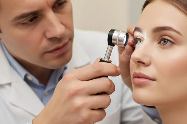
When you go to see a doctor, especially for a checkup or a routine checkup, you may have seen the doctor take a small flashlight-like device and look into your eyes, right? Some people think they are checking to see if your eyes can see. But it is actually a much deeper and more important test. This test can not only check the health of your eyes, but also find clues about some diseases in your entire body. Today we are talking about this test called Fundoscopy.

What exactly is fundoscopy?

Simply put, Fundoscopy is when your doctor examines the back wall of your eye. This part of the eye is called the 'fundus' in medical terms. That's why this test is called Fundoscopy.

The instrument used for this is called an ophthalmoscope . That's why this test is also called an ophthalmoscopy . This is a small instrument with magnifying lenses and a small light. It can shine light into your eye and see the important parts inside your eye clearly and magnified.

The eyes are like a window not only to our soul, but also to our physical health. A fundoscopy examination can reveal a lot by looking inside through that window.

There are two main ways to do this test.

There are two main ways a doctor can perform this test. The equipment used is slightly different.

Test method Description
Direct Ophthalmoscopy This uses a small, hand-held device. The doctor sits in front of you, places the device close to your eye, and looks inside. This is the method that doctors usually use most of the time.
Indirect OphthalmoscopyIn this, the doctor uses a device that is worn on the head (similar to a headlight). In addition, he holds another lens in front of your eye. This method allows you to see a wider area inside the eye, greatly magnified, and in three dimensions (3D). Ophthalmologists use this method more often.

When is a fundoscopy performed?

This is a very common test. Here are some situations where you might have to do this:

  • During a general medical examination (Physical exam): When you go for a general checkup, the doctor may do this.
  • When to see a pediatrician: This is a must-do when it comes to health check-ups for young children.
  • Eye exam: This is a must when you see an ophthalmologist for eye problems or eyeglasses.
  • Neurological exam: This exam is very important in cases of brain and nerve-related conditions.
  • For people with diseases like diabetes and high blood pressure: This is why doctors recommend that people with these diseases have their eyes checked at regular intervals.

What happens before and during the test?

There is nothing to be afraid of about this test. It is very simple and painless.

Before the test

Most of the time, you won't need to do any special preparation. However, sometimes, especially in an eye clinic, if the doctor wants to see inside your eye better, they may put some eye drops in your eye to dilate the pupil.

After using these drops, your eyelids will become larger, so you may be a little sensitive to light. Your vision may also be a little blurry. This may last for a few hours.

Important: If you have eye drops that make your eyes bigger, avoid driving for that day. If possible, have someone else with you. Wearing sunglasses will also help your eyes.

During the test

The doctor will ask you to sit in a chair or lie down on a bed. This test is usually done with the lights in the room dimmed so that you can see the inside of your eye clearly.

You will be asked to look straight ahead, and sometimes you may be asked to look in different directions. The doctor will take his ophthalmoscope and look into your eye. This will take a few minutes. You will not feel any pain during this.

What does this test look at inside the eye?

The doctor doesn't just look inside your eye. There are a few things he'll pay special attention to:

  • Retina: This is the light-sensitive membrane inside the eye. It is like the film in a camera. It is checked for any damage, bleeding, or other changes.
  • Optic Nerve: This is the 'wire' that carries messages from the eye to the brain. The base of this nerve (optic disc) is checked for swelling or discoloration.
  • Blood vessels inside the eye: By looking at the tiny blood vessels inside the eye, you can get an idea of ​​the condition of the blood vessels throughout the body.
  • Pupil response: We also pay attention to how your eyes respond to light.

What do the test results say?

This is the most important part. Fundoscopy can reveal a lot of information not only about eye diseases, but also about other diseases in the body.

Imagine, your doctor might find things like this with this test:

  • Diabetes: Diabetes can damage the small blood vessels in the eye. This can cause small hemorrhages or fluid accumulations (exudates) to appear in the eye . This condition is called Diabetic Retinopathy .
  • High Blood Pressure: High blood pressure can cause the blood vessels inside the eye to become thin, twisted, and stiff. Sometimes, this can also cause bleeding.
  • Increased Intracranial Pressure: When pressure inside the brain increases due to a brain tumor, hemorrhage, or other cause, the optic nerve root swells. This condition is called papilloedema . This can be easily detected with a fundoscopy.
  • Cataracts: This test can also detect clouding of the lens of the eye to some extent.
  • Glaucoma: This test can also detect damage to the optic nerve caused by increased eye pressure.

After the exam, the doctor will explain what he or she saw to you. If there are any problems, they may recommend further tests, such as a fundus photography (taking a picture of the inside of the eye) or a slit lamp exam , to find the cause. Don't be afraid to ask your doctor any questions you may have.

Take-Home Message

  • Fundoscopy (Ophthalmoscopy) is a painless, simple, but very important medical examination.
  • This can provide important information not only about the health of your eyes, but also about diabetes, high blood pressure, and some brain problems.
  • Don't be afraid of the doctor looking at your eyes during a routine medical exam. It's very important for your health.
  • If you use eye drops to enlarge your blackheads, your vision may be blurred for a few hours, so avoid driving and take someone with you to get home safely.
  • If you have any questions about your test or results, never hesitate to discuss them with your doctor.

Fundoscopy, Ophthalmoscopy, Eye Examination, Retina, Optic Nerve, Diabetic Eyes, Hypertensive Eyes
⚠️ Important: The medical articles and information on Nirogi Lanka are for general awareness only, and are by no means a substitute for professional medical advice, diagnosis, or treatment. For any medical problem you have, consult a qualified physician immediately.

💬 Comments (0)

No comments have been posted yet. Add your comment here for the first time.

Add your comment

Please calculate: 7 + 7 =
Why does the doctor look inside the eyes? Let's learn about Fundoscopy (Ophthalmoscopy) in simple terms!

Why does the doctor look inside the eyes? Let's learn about Fundoscopy (Ophthalmoscopy) in simple terms!

When you go to see a doctor, especially for a checkup or a routine checkup, you may have seen the doctor take a small flashlight-like device and look into your eyes, right? Some people think they are checking to see if your eyes can see. But it is actually a much deeper and more important test. This test can not only check the health of your eyes, but also find clues about some diseases in your entire body. Today we are talking about this test called Fundoscopy.

What exactly is fundoscopy?

Simply put, Fundoscopy is when your doctor examines the back wall of your eye. This part of the eye is called the 'fundus' in medical terms. That's why this test is called Fundoscopy.

The instrument used for this is called an ophthalmoscope . That's why this test is also called an ophthalmoscopy . This is a small instrument with magnifying lenses and a small light. It can shine light into your eye and see the important parts inside your eye clearly and magnified.

The eyes are like a window not only to our soul, but also to our physical health. A fundoscopy examination can reveal a lot by looking inside through that window.

There are two main ways to do this test.

There are two main ways a doctor can perform this test. The equipment used is slightly different.

Test method Description
Direct Ophthalmoscopy This uses a small, hand-held device. The doctor sits in front of you, places the device close to your eye, and looks inside. This is the method that doctors usually use most of the time.
Indirect OphthalmoscopyIn this, the doctor uses a device that is worn on the head (similar to a headlight). In addition, he holds another lens in front of your eye. This method allows you to see a wider area inside the eye, greatly magnified, and in three dimensions (3D). Ophthalmologists use this method more often.

When is a fundoscopy performed?

This is a very common test. Here are some situations where you might have to do this:

  • During a general medical examination (Physical exam): When you go for a general checkup, the doctor may do this.
  • When to see a pediatrician: This is a must-do when it comes to health check-ups for young children.
  • Eye exam: This is a must when you see an ophthalmologist for eye problems or eyeglasses.
  • Neurological exam: This exam is very important in cases of brain and nerve-related conditions.
  • For people with diseases like diabetes and high blood pressure: This is why doctors recommend that people with these diseases have their eyes checked at regular intervals.

What happens before and during the test?

There is nothing to be afraid of about this test. It is very simple and painless.

Before the test

Most of the time, you won't need to do any special preparation. However, sometimes, especially in an eye clinic, if the doctor wants to see inside your eye better, they may put some eye drops in your eye to dilate the pupil.

After using these drops, your eyelids will become larger, so you may be a little sensitive to light. Your vision may also be a little blurry. This may last for a few hours.

Important: If you have eye drops that make your eyes bigger, avoid driving for that day. If possible, have someone else with you. Wearing sunglasses will also help your eyes.

During the test

The doctor will ask you to sit in a chair or lie down on a bed. This test is usually done with the lights in the room dimmed so that you can see the inside of your eye clearly.

You will be asked to look straight ahead, and sometimes you may be asked to look in different directions. The doctor will take his ophthalmoscope and look into your eye. This will take a few minutes. You will not feel any pain during this.

What does this test look at inside the eye?

The doctor doesn't just look inside your eye. There are a few things he'll pay special attention to:

  • Retina: This is the light-sensitive membrane inside the eye. It is like the film in a camera. It is checked for any damage, bleeding, or other changes.
  • Optic Nerve: This is the 'wire' that carries messages from the eye to the brain. The base of this nerve (optic disc) is checked for swelling or discoloration.
  • Blood vessels inside the eye: By looking at the tiny blood vessels inside the eye, you can get an idea of ​​the condition of the blood vessels throughout the body.
  • Pupil response: We also pay attention to how your eyes respond to light.

What do the test results say?

This is the most important part. Fundoscopy can reveal a lot of information not only about eye diseases, but also about other diseases in the body.

Imagine, your doctor might find things like this with this test:

  • Diabetes: Diabetes can damage the small blood vessels in the eye. This can cause small hemorrhages or fluid accumulations (exudates) to appear in the eye . This condition is called Diabetic Retinopathy .
  • High Blood Pressure: High blood pressure can cause the blood vessels inside the eye to become thin, twisted, and stiff. Sometimes, this can also cause bleeding.
  • Increased Intracranial Pressure: When pressure inside the brain increases due to a brain tumor, hemorrhage, or other cause, the optic nerve root swells. This condition is called papilloedema . This can be easily detected with a fundoscopy.
  • Cataracts: This test can also detect clouding of the lens of the eye to some extent.
  • Glaucoma: This test can also detect damage to the optic nerve caused by increased eye pressure.

After the exam, the doctor will explain what he or she saw to you. If there are any problems, they may recommend further tests, such as a fundus photography (taking a picture of the inside of the eye) or a slit lamp exam , to find the cause. Don't be afraid to ask your doctor any questions you may have.

Take-Home Message

  • Fundoscopy (Ophthalmoscopy) is a painless, simple, but very important medical examination.
  • This can provide important information not only about the health of your eyes, but also about diabetes, high blood pressure, and some brain problems.
  • Don't be afraid of the doctor looking at your eyes during a routine medical exam. It's very important for your health.
  • If you use eye drops to enlarge your blackheads, your vision may be blurred for a few hours, so avoid driving and take someone with you to get home safely.
  • If you have any questions about your test or results, never hesitate to discuss them with your doctor.

Fundoscopy, Ophthalmoscopy, Eye Examination, Retina, Optic Nerve, Diabetic Eyes, Hypertensive Eyes
⚠️ Important: The medical articles and information on Nirogi Lanka are for general awareness only, and are by no means a substitute for professional medical advice, diagnosis, or treatment. For any medical problem you have, consult a qualified physician immediately.

💬 Comments (0)

No comments have been posted yet. Add your comment here for the first time.

Add your comment

Please calculate: 7 + 7 =