

Have you ever heard of an MRI scan? Maybe your doctor has asked you to have one. Or maybe a friend or relative has had one. So, you probably have a lot of questions about what an MRI is, why it is done, and what happens after it. Don't worry, today we will talk about this MRI (Magnetic Resonance Imaging) test in a simple way that you can understand.
What exactly is MRI (Magnetic Resonance Imaging)?
Simply put, an MRI is a painless test that allows us to see the inside of our bodies, such as organs and tissues, very clearly . It uses a large magnet, radio waves, and a computer to create detailed pictures of the inside of our bodies. The most important thing is that it does not use X-rays, or radiation.
Often, if someone needs to have frequent scans, especially for areas like the brain, doctors choose this MRI. The reason for this is that since there is no radiation, it does not cause much harm if done frequently. Imagine, when treating some diseases, you have to keep scanning to see if the disease is getting worse or better. In such cases, MRI is very useful.
What is an 'Open MRI'?
Now, there are two types of MRI machines. One is the 'Open MRI' type. In this, you lie down on a bed, and there are two flat magnets above and below you. This creates a large space on both sides. Some people feel claustrophobic when they go into the other type of MRI machine. For them, the 'Open MRI' is very good because it is less cramped.
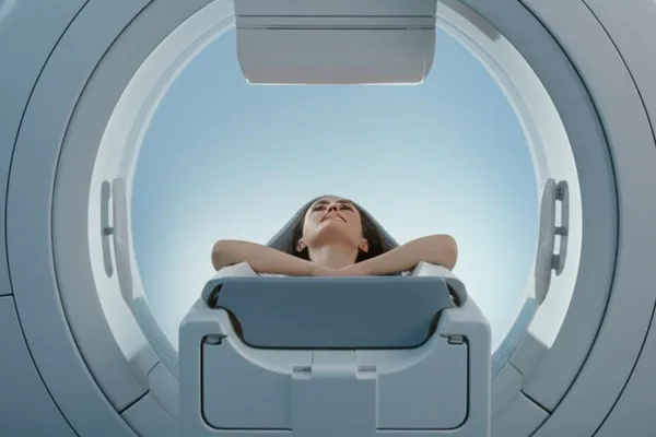
But there is a small problem. The images taken with this 'Open MRI' machine are not as clear as those taken with the other 'Closed-bore MRI' machines. The 'Closed-bore MRI' machine has a circle with magnets all around it. It's like a tube. You go inside that tube. So it's a little narrow, and there's not much space between your head and the part of the machine above. Because of this, some people may feel a little uncomfortable and scared. However, the best quality images can be taken with this machine.
If you are having an MRI scan and you are a little afraid of tight spaces, talk to your doctor about it. He or she will be able to give you sedatives or even anesthesia if needed.
What is an MRI with 'contrast'?
During some MRI scans, you are given a small injection of contrast material. It contains a rare metal called gadolinium. When this material is injected into your body, it changes the magnetic properties of nearby water molecules. This makes the images clearer . This increases the sensitivity and specificity of the image.
Using this 'contrast' material increases the clarity of these things:
- Tumors
- Inflammatory conditions (`Inflammation`)
- Infection
- How blood is supplied to certain organs
- Blood vessels
If your MRI requires this type of contrast material, a healthcare provider will insert a small cannula (an ``intravenous catheter (IV line)'') into a vein in your arm or hand. This is where the medicine is injected.
This 'contrast' material is a safe medicine . Sometimes minor side effects can occur, but serious reactions are very rare.
What is the difference between an MRI and a CT scan?
MRI (Magnetic Resonance Imaging) uses magnets, radio waves, and a computer to create images of the inside of the body. However, CT (Computed Tomography) scans use X-rays and computers.
Doctors often choose an MRI over a CT scan to look at soft tissues , which are non-bony parts of the body. MRI scans are also safer because they do not contain the ionizing radiation that X-rays do.
Not only that, but an MRI can see things like your brain, spinal cord, nerves, muscles, ligaments, and tendons much more clearly than regular X-rays and CT scans.
However, not everyone can have an MRI. The strong magnetic field of an MRI can move some metal implants inside the body, or affect the functioning of devices like pacemakers and insulin pumps. In such cases, a CT scan is the next best option.
An MRI scan is usually a little more expensive than an X-ray or CT scan.
What can be checked with an MRI?
We've heard that MRI (Magnetic Resonance Imaging) can take detailed pictures of the inside of your body. So, doctors can "see" many things inside your body with this MRI, including:
- Your brain and the nervous tissue around it .
- Organs in the chest and abdomen. Examples include the heart, liver, biliary tract, kidneys, spleen, bowel, pancreas, and adrenal glands.
- Breast tissue.
- Your spine and spinal cord .
- Pelvic organs. These include the bladder and reproductive organs (uterus and ovaries in women, prostate gland in men).
- Blood vessels.
- Lymph nodes.
When do I need an MRI?
Doctors use MRI scans to diagnose various medical conditions and to see how well treatments are working. There are different types of MRIs, depending on what part of your body you want to see.
Brain and spinal cord MRIs help diagnose and treat conditions such as:
- Brain aneurysms.
- Brain tumors and spinal tumors.
- Damage to the brain or spine due to an accident.
- Compression or inflammation of the spinal cord and nerves (e.g., `pinched nerve`).
- Multiple sclerosis (MS).
- Spinal cord-related conditions.
- The structure and position of the spine.
- Stroke.
Cardiac (heart) MRIs are used for several reasons:
- Look at the size of your heart chambers, heart valves, major blood vessels and how blood flows through them, as well as the shape and function of surrounding structures.
- Identify cardiovascular conditions, such as tumors, infections, and inflammatory conditions.
- Assess the effects of coronary artery disease. For example, restricted blood flow to the heart muscle, scarring of the heart muscle after a heart attack.
- See the structure and function of the heart and blood vessels in children and adults with congenital heart disease.
MRIs for other parts of the body (Body MRIs) can look at structures and conditions such as:
- Lumps in your chest, abdomen, or pelvis.
- Liver diseases, such as cirrhosis, as well as problems with the bile ducts and pancreas.
- Inflammatory bowel disease, for example, Crohn's disease and ulcerative colitis.
- Blood vessel abnormalities and inflammation of blood vessels (vasculitis).
- The developing fetus in the womb.
MRIs of bones and joints help to check for:
- Bone infection (osteomyelitis).
- Bone tumors.
- Disc abnormalities in the discs between the vertebrae of the spine.
- Joint problems due to injuries.
Sometimes doctors do breast MRIs along with mammography to detect breast cancer, especially in women with dense breast tissue or those at high risk of developing breast cancer.
Is it safe to have an MRI?
In general, if proper safety precautions are followed , an MRI scan is a very safe procedure. It poses no risk to the average person.
The strong magnetic field emitted by the MRI machine is not harmful to you. However, it may cause some implanted medical devices inside your body to not work properly or may distort the images.
If your MRI uses contrast material, you may very rarely have an allergic reaction. These reactions are usually mild and can be controlled with medication. If you have an allergic reaction, a healthcare professional will be there to help you right away.
Pregnant women are not allowed to have MRIs with gadolinium contrast unless absolutely necessary , as the risks to the fetus are not fully known.
Who should not have an MRI?
In most cases, it is safe for people with metal implants to have an MRI. However, some types are not . If the device you have is not certified as MRI-safe, you may not be able to have an MRI. Such devices include:
- Metallic joint prostheses.
- Some cochlear implants.
- Some types of clips used for cerebral aneurysms.
- Some types of metal coils inside blood vessels.
- Some older cardiac defibrillators and pacemakers.
- Vagal nerve stimulators.
If your doctor asks you to have an MRI, he or she will ask about your medical history and any medical devices you have inside or outside your body. It's very important to be accurate about all of this.
Who does an MRI?
Your MRI will be performed by a radiologist or a radiology technologist. A radiologist is a doctor who examines and analyzes these images to diagnose diseases. A radiology technologist is a healthcare professional who is specially trained and certified to perform MRI scans.
How does the MRI machine work?
Here's how an MRI (Magnetic Resonance Imaging) machine works: An electric current is passed through a coil of wire, creating a temporary magnetic field in your body. Then a transmitter/receiver in the machine sends and receives radio waves. A computer uses these signals to create digital images of the area being scanned. Isn't it amazing technology?
How should I prepare before an MRI?
Because the MRI scanner uses strong magnets and radio waves, some metal objects in your body may heat up or move around. This can cause health and safety issues. It can also cause some electronic medical devices inside your body to malfunction.
Important: If you have any metal objects or implanted medical devices in your body, you must tell your doctor and MRI technologist about them before the MRI.
Some implants may require additional planning and special instructions. Others may not require special instructions, but you may need to take an X-ray to see exactly where the implant is before the procedure.
Please tell your doctor and MRI technologist if you have any of these:
- Heart pacemaker or defibrillator.
- Electronic or implanted stimulators or devices. For example: deep brain stimulators, vagus nerve stimulators, bladder stimulators, spine stimulators, neurostimulators, and implanted electrodes or wires.
- Metallic joint prostheses.
- Cochlear implant or other ear implants.
- Implanted medication pumps. For example, those that pump narcotic/pain medications or medications for spasticity.
- Programmable shunt.
- Aneurysm clips and coils.
- Stents not located in your heart.
- Filters, e.g. blood clot filters.
- Metal fragments in your body or eyes. For example, bullets, shrapnel, metal fragments, or gunpowder.
You cannot wear these devices during an MRI. Try to wear them during the MRI on the day you change your patch or device:
- Continuous glucose monitor (CGM).
- Insulin pump.
- Medication patches.
In addition, tell your doctor about these things:
- "Are you pregnant ?"
- Is it difficult to lie on your back for 30 to 60 minutes?
- Do you have a fear of confined spaces (claustrophobia)?
Leave all jewelry and other accessories at home, or remove them before the MRI. Metal and electronic items are not allowed in the examination room. They can interfere with the magnetic field of the MRI machine, cause burns, or become `harmful projectiles`. Examples include:
- Jewelry, watches, credit cards, and hearing aids - they can all break.
- Pins, metal hairpins, underwired brassieres, and metal zippers - these can distort MRI images.
- Removable dentures.
- Pens, pocket knives and glasses.
- Body piercings.
- Mobile phones, electronic watches and tracking devices.
How long does an MRI scan take?
Depending on the type of test and the equipment used, the entire test usually takes between 30 and 50 minutes to complete. Depending on the reason for your scan, your doctor may be able to give you a more precise time.
What is the experience like when having an MRI?
Most MRI scans are painless . However, some people may find it difficult to remain still for 30 minutes or more. Others may feel intimidated by the feeling of being confined inside the MRI machine. The machine can make a little noise .
Here are some things that typically happen during an MRI scan and what you should expect:
- For the MRI scan, you will have to wear a hospital gown.
- In most tests, you will need to lie face up on the MRI scanning bed.
- The MRI scan bed is placed inside the MRI machine.
- When the MRI scan begins, you will hear a series of loud clicking and tapping sounds as the machine takes the images. This may continue for several minutes. Before the test begins, you will be given earplugs or headphones to protect your ears.
- It is very important to remain very still during the test to get the best quality images .
- It is normal to feel a slight warmth in the part of your body being scanned. If this bothers you, tell the radiologist or technologist.
- The MRI technologist can see you at all times and can talk to you. There is a two-way intercom system while you are inside the scanner. You also have a call button on your hand. If you have any questions or concerns, you can let the technologist know.
In some cases, your MRI may require contrast material. If this is the case, a healthcare provider will give you the contrast material through an IV before the MRI. You may feel some pain from the IV needle, but it is not severe. You may also experience some bruising afterward. Some people may experience a temporary metallic taste in their mouth after the contrast material is injected.
If you have claustrophobia, your doctor may give you a sedative or anesthesia to help you stay calm during the test.
Are there any side effects of MRI 'contrast'?
Very rarely , some people who have had contrast material used for MRI may experience side effects such as:
- Nausea.
- Headache .
- Pain at the site of the injection.
Allergic reactions to contrast material, such as hives and itchy eyes, are very rare . If you experience any allergic symptoms, tell the technologist. A health worker will be there to provide immediate medical attention.
People with kidney disease may develop a rare complication called nephrogenic systemic fibrosis (NSF), which causes thickening of the skin, organs, and other tissues when they have an MRI with contrast. Therefore, people with severe kidney disease may not be able to have gadolinium-based contrast agents for MRI.
There is evidence that after an MRI with contrast, a very small amount of gadolinium can remain in various organs in your body. Although this has no known adverse effects, your doctor may consider this `gadolinium retention` when choosing a contrast agent.
What happens after an MRI?
Unless you took a sedative drug for your MRI scan, you don't need any special recovery time. You can go home and do your normal activities. However, if you took a sedative drug for the test, you will need to recover from the effects of the drug before you go home. You may need someone to drive you home.
When will I know the MRI results?
After your MRI scan, a radiologist will analyze the images. The radiologist will send a signed report to your primary care provider, your doctor. He or she will tell you the results. You may need to have another test (a follow-up exam). If so, your doctor will explain why.
Remember as a summary (Take-Home Message)
MRI (Magnetic Resonance Imaging) is a very useful and generally safe imaging test that doctors use for a variety of reasons. If you are having an MRI scan and have any concerns or questions about the test, don't be afraid to ask your doctor. They are there to help and support you.
Remember, an MRI is not an X-ray. It uses a strong magnetic field and radio waves. So there is no radiation. However, if you have any metal objects in your body, it is imperative to tell your doctor about it.
So, the next time you hear about an MRI, you now have a better understanding of it, right?
` MRI, MRI, scan, magnetic resonance imaging, diagnosis, health tests, medical technology




💬 Comments (0)
No comments yet. Be the first to share your thoughts here.
Add Your Comment