

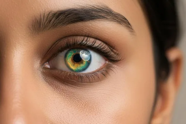
Have you ever wondered how we see the world so clearly? The main reason for this is the transparent, thin membrane in front of our eyes. This part, which works like the lens of a camera, is called the "Cornea". If the shape of this cornea changes even slightly, what we see can become blurry and distorted. In such cases, we call it `Corneal Topography`, a very important and painless test to check the exact shape of your cornea.
Simply put, what is Corneal Topography?
This name may sound a bit strange. We usually use the word "topography" in geography, which is to make maps that show the surface of the earth, the shape of mountains, valleys, rivers, etc. So how do you visually draw a map?
Imagine that your cornea is like a small island. Some parts of the surface of this island are high, some parts are low, and some parts may have holes. But we simply cannot see these changes with our eyes. That is why a technique called `Corneal Topography` is used. What it does is, it uses a special computer device to create a three-dimensional (3D) map of your cornea. This map is displayed in color. With those colors, we can see exactly where the cornea is high, where it is flat, where it is thin, and where it is thick.
Simply put, this is like a special photo shoot for your eyes. There is no pain, and nothing touches your eyes.
Why do we need this test?
This test serves several very important functions in ophthalmology. There are three main reasons.
1. Identify eye diseases and monitor their progress
Some eye diseases directly affect the shape and thickness of the cornea. This test is essential for diagnosing such diseases.
- Astigmatism: A condition in which the cornea is not perfectly round, but rather slightly elongated, like an egg. This can cause blurred vision.
- Keratoconus: This is a somewhat serious condition. What happens here is that the cornea gradually thins and bulges forward like a cone. A `Corneal Topography` test is very important to detect this at an early stage.
- Pellucid marginal degeneration (PMD): This is also a condition in which the cornea thins.
- Scars caused by injury or infection: If there is a scar on the cornea, you can tell exactly how it affects vision.
- Pterygium: A condition in which a fleshy growth grows from the white of the eye onto the cornea.
2. Plan surgeries and check results
This test is essential in eye surgeries, especially corneal surgeries.
- Laser vision correction surgery (LASIK or PRK): Many people want to get rid of glasses or contact lenses by having `(LASIK)` surgery. But not everyone can have this surgery. This test is definitely done to make sure 100% that the shape and thickness of your cornea are suitable for this surgery. As mentioned earlier, a condition like `(Keratoconus)` can be detected even if it is asymptomatic. If `(LASIK)` is done for such people, serious complications can occur.
- Cataract surgery: When removing a cataract and implanting an artificial lens, information about the shape of the cornea is important to accurately calculate the power of that lens.
- Cornea transplant surgery: This test helps to check the condition of a damaged cornea before and after it is replaced with a new one.
- Corneal cross-linking: A surgery to strengthen the cornea that has become weakened due to diseases such as `(Keratoconus)`. This test is also necessary to plan this surgery.
3. Choose contact lenses that fit your eyes perfectly.
This test is not always necessary to fit a regular contact lens. However, if your cornea is abnormally shaped, such as Keratoconus, regular lenses will not work. In that case, you will need to use special rigid contact lenses (Rigid gas-permeable - RGP lenses) . In order for such a lens to fit your eye perfectly, all the measurements of the cornea must be taken and the lens must be made to fit it. The map obtained from Corneal Topography is very useful for this.
Who wants to take this test?
You may be wondering if you need this test. Doctors usually recommend this test in the following situations.
| Situation | Reason |
|---|---|
| If you are considering laser surgery (such as LASIK) | Make sure your cornea is suitable for surgery and that there are no hidden medical conditions. This is a mandatory step. |
| If you need surgery for a corneal condition | It helps the doctor plan the surgery precisely and achieve the best results. |
| If you have already had eye surgery | Check the results of the surgery and how the shape of the cornea has changed. |
| If you need special contact lenses | Choose a comfortable lens that fits perfectly for an unusually shaped cornea. |
The test is very easy!
Do you need to prepare before the test?
Usually, no special preparation is needed. However , if you wear contact lenses, your doctor will tell you to stop wearing them a few days, or even weeks, before the test. This is because wearing lenses can temporarily change the natural shape of your cornea (corneal warpage). So to get accurate measurements, you need to wait until your cornea has returned to its natural shape.
What happens during the test?
This entire process only takes a few minutes.
1. You are made to sit on a chair in front of a machine that looks like a big ball.
2. Your forehead and chin are placed in the designated areas and are told to stay comfortable.
3. Then they are asked to stare straight at a certain light inside the ball.
4. While you wait, the machine takes several pictures of your eyes without any noise or pain.
5. The images taken are analyzed by a computer and converted into the aforementioned color maps.
That's it! You don't feel anything. The machine doesn't even hit your eyes . It's just like taking a photo.
The results of the test... colored maps!
After the test, your doctor will give you several colored maps. When you see these, you may not be able to figure out what they are. But each color and each number has a meaning.
Generally, warm colors like red and orange indicate high/protruding areas of the cornea . Cool colors like blue and green indicate flat/low areas of the cornea .
Depending on the information the doctor needs, different types of maps can be obtained.
| Map Type | What does this show us? |
|---|---|
| Axial Map (Sagittal Map) | This is the most commonly used map. It shows the curvature of the cornea, i.e., the high and low points (like hills and valleys). In a healthy cornea, this map is often green. |
| Elevation Map | This compares your cornea to a computer-generated "standard shape." Warm colors indicate areas that are higher than the standard shape. Cold colors indicate areas that are lower than the standard shape. |
| Corneal Thickness Map (Pachymetry map) | This map shows the thickness of the cornea. Warm colors (like red) indicate thinner areas, and cool colors (like blue) indicate thicker areas. This map is important for diagnosing conditions like keratoconus, where the cornea becomes thinner. |
Don't be alarmed by these colorful maps. It is your doctor's responsibility to accurately analyze the information on these maps and provide an explanation for your condition. Ask him or her any questions you may have.
Is this safe? When should I talk to the doctor?
Yes. Corneal Topography is a 100% safe test. There are no known risks or side effects. It is also a painless, quick test.
If you have any questions about your test results, or if your eye symptoms (such as blurred vision, pain) are new or worsening, see your doctor right away.
Take-Home Message
- Corneal Topography is a painless and safe test that creates a 3D map of the cornea (the transparent part at the front of your eye).
- This is essential to diagnose diseases such as `Keratoconus` and `Astigmatism`, and to determine whether you are suitable for laser surgery such as `LASIK`.
- This helps create specialized contact lenses for abnormally shaped corneas.
- The test results are presented in the form of color maps. Your doctor will explain the meaning of these to you in simple terms.
- If you have any questions about this test or the results, don't be afraid to ask your doctor.




💬 Comments (0)
No comments yet. Be the first to share your thoughts here.
Add Your Comment