
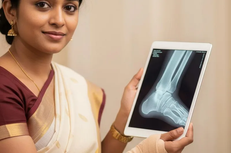
Maybe you've twisted your leg while walking, running, or playing. Or maybe you've had unbearable pain or swelling in your leg or ankle for no apparent reason. When you went to the doctor, did he tell you to get an X-ray of your leg? Some people get a little scared when they hear the word 'X-ray'. They think things like, "What will happen with this?", "Isn't radiation good for the body?" But it's actually a very simple, painless test that usually takes just a few minutes. Let's talk about this from the beginning today.
What exactly does an X-ray of this leg mean?
Simply put, a foot X-ray is a black-and-white picture of the inside of your foot. It's like a photograph. This picture shows the bones of your foot, including the tarsal bones, metatarsal bones, and phalanges, as well as some of the soft tissues. Doctors use this picture to help diagnose the problem with your foot or ankle. This is sometimes called a 'foot radiograph'.
So how does this X-ray machine work?
This is very interesting. An X-ray machine sends a special type of light that our eyes cannot see, called electromagnetic waves, through our bodies. Imagine you are sitting in a dark room and you shine a flashlight in front of your hand. Can you see that the light is less reflected from the bones of your hand, which means there is more shadow? An X-ray is like that.
Different parts of our body absorb these X-rays to different degrees.
- Bones: Because our bones are rich in calcium, they absorb a lot of X-rays. That's why bones appear white on an X-ray.
- Soft tissue: Soft tissues such as muscle, fat, and organs absorb less radiation, so they appear as varying shades of gray .
- Air: Because rays pass through air without any obstruction, those areas appear black .
By looking at the image obtained in this way, a doctor can easily detect things like a bone fracture, a fracture, or a dislocated joint.
In what cases is a leg X-ray taken?
If you have unexplained pain, swelling, or tenderness in your leg or ankle, your doctor may recommend an X-ray to find the cause. This is also important to check for damage to your leg after an accident.
Let's look at the table below to see the main situations in which an X-ray is used.
| Condition or cause | A simple explanation |
|---|---|
| Fractures | Check to see if there is a broken bone or a fracture in the leg or ankle. |
| Dislocated joints | Check to see if the joint, where two bones meet, has shifted. |
| Joint deformities (e.g. bunions) | Some people experience pain when the joint of their big toe protrudes forward. Identify this condition as ``hallux valgus``. |
| Conditions such as arthritis | Understand the causes and severity of joint pain that occurs with aging or other causes. |
| Cysts | Identify abnormal fluid-filled cysts that form in the bones. |
| Bone infections | Check for bone infections caused by bacteria. |
| Bone cancer | Although this is very rare, X-rays can help detect cancerous conditions in the bones. |
| Post-treatment check-up | If a cast is placed after a broken leg, an X-ray will also be taken to see if the bones are properly set and healing. |
How should I prepare before an X-ray?
A leg X-ray doesn't require much preparation, but there are a few important things to keep in mind.
- Clothing and Jewelry: When you go for an X-ray, you will need to remove any metal objects, jewelry, shoes, or socks on or near your leg. These can interfere with the clarity of the X-ray image. Therefore, it is best to wear loose clothing that can be easily removed.
- Pregnancy (very important): If you are pregnant or have any suspicion that you may be pregnant, be sure to tell the radiologic technologist who is taking the X-ray. This is very important. This is because the radiation exposure to the developing baby needs to be minimized. The doctor will only decide to do the X-ray if absolutely necessary, and with special safety precautions.
Before the X-ray is taken, the technician in the room will explain everything to you. Any questions or concerns you may have can be addressed at that time.
What happens during the X-ray?
This process is very quick, usually taking less than 15 minutes.
1. Entering the room: You will be taken to a special room where the X-ray will be taken. These rooms are usually a bit cool.
2. Protective clothing: To protect your reproductive organs from radiation, you can wear a heavy garment containing lead, such as a lead apron, on your lap.
3. Positioning: Next, the technician will ask you to position your leg in the correct position on the X-ray table. You may also be asked to place pillows or sandbags on either side of your leg to help keep it in place. It is very important to remain still during the X-ray. If you move, the image may be blurry.
4. Taking pictures: The technician will then go behind a special wall or into a separate room and turn on the X-ray machine. You will not feel anything during this time. The pictures are usually not taken in the same position. To get the best view, the leg is turned in different directions (front, side, at a 45-degree angle) and about three separate pictures are taken.
It's like looking at a house from just one side and not seeing everything inside. You have to look at it from different angles to find the exact location of the problem.
If you feel any pain at this time, tell the technician. He or she will help you to be as comfortable as possible.
What happens after the X-ray is taken?
After the X-ray images are taken, the technician will ask you to wait a few minutes to check if the images are clear or blurry. If any images are unclear, they will be retaken immediately.
These images are then sent to a specialist.
- Radiologist: A radiologist is a doctor who has received special training to diagnose diseases by examining images such as X-rays and CT scans and accurately analyzing their contents.
- Report: He looks at these pictures, includes his observations, and creates a report.
- Your doctor: This report will be given to your treating doctor. The doctor will then explain to you what your condition is and what treatment is needed next, based on the report.
Sometimes, the doctor may need to take another X-ray if they want to see the leg from more angles. Or, they may recommend another X-ray to see how things are after treatment.
Is there any risk in this?
This is something that many people are afraid of. Since X-rays use radiation, the question arises as to whether it is good for the body or not.
True, X-rays use a small amount of radiation. But that amount is very small. Simply put, the amount of radiation you are exposed to from an X-ray of one leg is about the same as the amount of natural radiation your body is exposed to when you live in a normal environment for a few days. So it won't cause much harm.
Doctors say that the benefits of getting an accurate diagnosis far outweigh the small radiation risks . Not knowing exactly what the illness is is even more dangerous.
- Pregnant women and children: As mentioned earlier, pregnant women and young children are at increased risk from radiation, so special care is taken with them. If children are X-rayed, the lowest possible dose of radiation is used.
There is a small risk of cancer from very high levels of radiation exposure. But a single X-ray is unlikely to cause that risk. If you have any further questions about this, ask the technician taking the X-ray or your doctor.
How long does it take to receive the report?
If you have been admitted to the Emergency Department (ETU) of a hospital after an accident, you can get the X-ray results almost immediately. Normally, it can take a day or two for the radiologist's report to reach your doctor. The doctor will then discuss the results with you.
Take-Home Message
- A leg X-ray is a very simple, quick, and painless test. There's no reason to be afraid of it.
- This mainly looks at the condition of the bones in your leg. This helps to accurately determine if there is a fracture, dislocation, or other problem.
- If you are pregnant or suspect you may be pregnant, be sure to inform the staff before having the X-ray.
- The amount of radiation from an X-ray is very small . The benefits of obtaining an accurate diagnosis far outweigh the small risk.
- Your doctor will explain the X-ray results and the next steps to take.




💬 Comments (0)
No comments yet. Be the first to share your thoughts here.
Add Your Comment