

Have you ever heard an eye doctor say, "Let's take a picture of the inside of your eye"? You might have been a little surprised when you heard that. A picture of the inside of your eye? How do you take one? What do you see in it? Many questions must have come to mind. The medical name for it is Fundus Photography . This is actually a painless, very simple and important test to check the health of your eye. So, let's talk about this clearly and simply today.
What exactly is Fundus Photography?
Simply put, Fundus Photography is a procedure that takes a picture (photo) of the back wall of your eye, called the fundus . This photo allows your eye doctor to clearly examine some of the most important parts of your eye that are essential to your vision. Just like taking an X-ray of the inside of our body, this allows us to see the inside of our eye.
Think of our eye as a camera. Light from outside enters the eye and hits a screen at the back. That screen-like part is what this test mainly looks at. The main parts of that area are:
- Retina: This is the layer of light-sensitive cells (`photoreceptors`) at the back of the eye. It's like the film in a camera. These cells detect light and send that information as electrical signals to the brain. The brain combines those signals to create the image we "see."
- Macula: This is the most sensitive part of the retina. The light-sensitive cells in this part help us see color and fine details clearly. This is what helps you do fine things like read a book and recognize someone.
- Optic Nerve: This is like a data cable between our eyes and our brain. All the information gathered from the retina is carried to the brain through this nerve. Many eye conditions can affect this optic nerve.
- Choroid: This is the layer beneath the retina. It contains the network of blood vessels that supply the retina with blood, oxygen, and nutrients.
Keeping all of these parts healthy is very important for your vision. Fundus photography can detect even the slightest changes in these parts.
When is fundus photography done?
This test can also be done as part of a routine eye exam. This means that it is done to make sure that there are no problems with your eyes. In addition, if you have any eye symptoms or feel that your vision has changed, this test is definitely done to find the cause.
Fundus photography is especially important to check for the presence of the following diseases or to monitor whether those diseases are under control.
| Condition or cause | A simple explanation |
|---|---|
| Diabetic Retinopathy | Check to see if the blood vessels in the eye have been damaged or bleeding due to diabetes. This is very important for diabetics. |
| Glaucoma | Check for damage to the optic nerve due to increased eye pressure. |
| Macular Degeneration | Recognize changes in the macula that can occur with age and affect clear vision. |
| Retinal tears or detachments | Check for a torn, detached, or otherwise damaged retina. |
| Optic nerve conditions | Check for conditions such as swelling of the optic nerve (papilledema) or drying out (optic atrophy). |
| Tumors inside the eye | Check for cancerous (e.g., retinoblastoma) or noncancerous tumors inside the eye. |
| Scotomas (black spots in vision) | If you are losing some areas of your vision, find out where the cause is inside the eye. |
How does the machine that performs this test work?
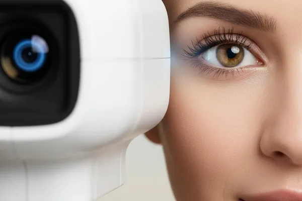
A special camera is used to perform fundus photography. This is called a `fundus camera`. Eye clinics usually have at least one of these types of cameras. There are mainly two types of cameras.
1. Standard fundus camera: This is a device that looks a bit like a pair of binoculars. It has a separate frame for you to place your chin and forehead on. There is a lens on the other side for the doctor to see. Before taking the photo with this type of camera , some pupil-dilating drops are put in your eye to make the pupil dilate. This allows you to see the inside of your eye clearly.
2. Wide-field laser-based cameras: These are relatively new cameras. They can take a clear, wide-angle photo of the inside of the eye without magnifying the pupil. However, sometimes the image becomes clearer when you put in eye drops. In this case, you have to look into a circular hole.
What to expect before and during the test?
You do not need to make any special preparations for this test.
However, if your doctor decides to put eye drops in your eye to make your blackhead bigger, it will take a few minutes (usually 15-20) for the medicine to work. During that time, you will have to wait a while.
During the test...
Your experience may vary slightly depending on the type of camera your doctor uses.
- If from a regular camera:
You will sit in a chair and place your chin and forehead in the frame of the camera. This will help you take a clear picture quickly because your head won't move. When you look into the device, you will see a small light. You will need to look at it. The doctor or technician on the other side will adjust the camera and take pictures one by one. This can be done for one or both eyes.
- If from the laser camera:
In this, you are asked to look straight at a circular target. As you look, a bright green light , like a photocopier light, passes through your vision for about a second. That's when the photo is taken. Usually, about three photos are taken with each eye.
This test is very quick. It only takes a few seconds to take a single photo. And it's completely painless .
What happens after the test? How will I know the results?
After the photos are taken, you will have the opportunity to talk to your doctor. Most cameras now allow you to view the photos directly on your computer screen.
The doctor will immediately put the photos on the computer screen and show them to you, explaining what's inside the eye.
- If everything is normal , the doctor will tell you so.
- If there are any changes or signs of illness , they will explain what they are and what to do about them.
- If you've had this test before, you can also compare new photos with old ones . This will help you see exactly what changes have occurred inside the eye over time.
Depending on the results of the photos, your doctor may do one or more of the following:
- Recommending some more tests.
- Suggesting treatment methods.
- Providing a date for a follow-up appointment.
- Referral to another specialist.
Are there any risks or side effects to this?
Fundus Photography is a very safe test . There are almost no side effects from this test.
However, if you have dilating drops to enlarge your black eye, you may experience some minor side effects. But don't worry, these are temporary .
- Vision may be a little blurry.
- It may be difficult to adjust the eyes to bright light (`light sensitivity` or `photophobia`).
This condition usually goes away completely within a few hours. Your doctor will tell you how long the effects of this medicine will last.
Can I drive after the test?
This is a problem that many people have. If you have had your black eye enlarged with eye drops, it is safest to avoid driving for a few hours . This is because your vision is blurred, which increases the risk of an accident. Therefore, it is better to come with someone or use public transportation when coming to this test than to drive alone.
The clinic will usually give you disposable shaded covers that can be worn like sunglasses to reduce sensitivity to light.
Take-Home Message
- Fundus Photography is a very simple and painless test that takes a picture of the inside of the back of your eye.
- This test can detect many conditions, such as diabetes, glaucoma, and macular degeneration, perhaps even before you have symptoms.
- The test is very safe. The only side effects are due to the eye drops that enlarge the black eye, and even those are temporary.
- If you put drops in your eyes, your vision may be blurred for a few hours. Therefore, it is wise to avoid driving for that day.
- If you have any questions about this test or the results, don't be afraid to ask your doctor. It's very important to be aware of your eye health.




💬 Comments (0)
No comments yet. Be the first to share your thoughts here.
Add Your Comment