
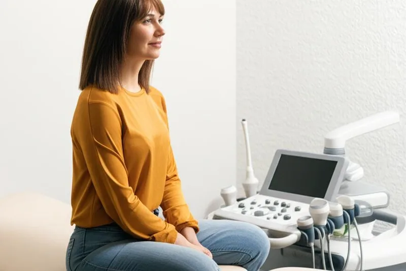
You may have been told by a doctor to have a liver ultrasound, right? Or you may have heard about it somewhere. It's actually a very simple, painless, and easy test to do. Doctors often recommend this test first to see if there is any problem with the liver. So, let's talk about this liver ultrasound in a clear and simple way, shall we?
Why would a doctor recommend a liver ultrasound?
Imagine that you have some symptoms related to liver disease, or that your doctor has noticed a slight change in the results of a blood test you did. In such a case, this ultrasound test is the easiest way to quickly find out if there is really something wrong with the liver. Also, doctors use this test to monitor whether the condition is under control and whether the liver is functioning well in someone who already has a liver disease. Simply put, this is the first and most important step in checking the liver.
What problems does an ultrasound show in the liver?
A liver ultrasound can detect various changes in our liver. For example:
- Steatotic Liver Disease - This is also called " fatty liver".
- Inflammation and swelling of the liver (Hepatitis)
- Whether scar tissue has formed in the liver (Fibrosis or Cirrhosis) - This is the risk of the liver gradually failing.
- Whether there are any abnormal spots or lumps in the liver (Liver Lesions).
These are the three main stages of long-term liver disease. In addition, special types of ultrasound can also be used to check things like the blood supply to the liver and the stiffness of the liver tissue.
What types of conditions can a liver ultrasound help diagnose?
A liver ultrasound is a very important tool in the diagnosis of many diseases. But remember, it alone may not be able to confirm every disease. However, it can be a great help to point the doctor in the right direction. Depending on the results of the ultrasound, further tests may be needed. For example, if you see a lump in your liver, you may be asked to do a biopsy to find out exactly what it is.
Here are some conditions that ultrasound can help diagnose:
- Stages of long-term liver disease:
- Hepatitis
- Steatosis (fat deposition)
- Cirrhosis
- Liver lumps/spots:
- Liver Cysts
- Liver Hemangioma (harmless tumor made up of blood vessels)
- Liver Cancer
- Conditions related to the blood vessels of the liver:
- Ischemia (reduced blood supply)
- Budd-Chiari Syndrome
- Portal hypertension (increased pressure in the vessels that carry blood to the liver)
- Other conditions that affect your biliary system:
- Ascites (fluid accumulation in the abdomen)
- Cholecystitis (inflammation of the gallbladder)
- Choledocholithiasis (Bile duct stones)
What are the types of liver ultrasound?
There are different types of liver ultrasound tests. A doctor will decide which type to perform based on the specific need. Let's look at a few of the main types.
1. Standard Ultrasound
This is often the first ultrasound done. It is sometimes called an "Upper Right Quadrant Abdominal Ultrasound" because the liver is located in that part of the abdomen. Although it mainly looks at the liver, it can also look at the gallbladder, bile ducts, right kidney, and part of the pancreas. Because all of these are interconnected.
2. Vascular Ultrasound / Doppler Ultrasound
This specifically looks at the blood vessels inside the liver and how blood flows through them. This is also called a Doppler Ultrasound . Along with regular ultrasound images, Doppler technology also shows the speed and direction of blood cells moving. Therefore, it can be used to better determine if there are any problems with the veins that supply blood to the liver.
3. Elastography Ultrasound
This is a special type of ultrasound. It is also called transient elastography . It measures the degree of stiffness, or fibrosis, in the liver. Fibrosis occurs when scar tissue builds up in the liver. If it gets too bad, it can lead to cirrhosis . In this test, a special device called a transducer sends shock waves through the liver. The speed at which the waves travel determines the level of fibrosis. The faster the speed, the greater the fibrosis.
4. Contrast-Enhanced Ultrasound (CEUS)
If a liver lesion is seen during a regular ultrasound, a doctor may order a CEUS test to study it more clearly. In this test , a contrast agent is injected into a vein. However, this is not a dye, but tiny gas bubbles.Some. When these go to the liver through the blood, those spots are clearly visible. These gas bubbles do not cause any harm to the body, and the chance of an allergic reaction is very low.
How should I prepare for a liver ultrasound?
One advantage of this test is that it doesn't require much preparation . However, if possible, your doctor will ask you to fast for 6 to 12 hours . This will empty your digestive tract and reduce the amount of food and gas that can interfere with the ultrasound waves. The gallbladder will also be enlarged and clearly visible.
Be sure to ask your doctor about drinking water. Some people also recommend stopping drinking water a few hours before the scan. Sometimes a full bladder can help with the scan.
What happens during a liver ultrasound?
You will have this test in a hospital or a specialized imaging center . You will be asked to lie on an examination table, often with your arm facing up. The ultrasound machine has a computer screen and a small wand-like device called a transducer wand. This transducer sends high-frequency sound waves that we cannot hear into your liver.
Then, the sonographer applies a clear, hypoallergenic gel to your abdomen. This gel creates a good contact between the transducer and your skin, without any gaps. Then, he or she moves the transducer back and forth over the gel. You won't feel any pressure or pain, unless you have severe pain in the area. As the transducer is moved, images of the liver and surrounding organs are displayed on the computer screen in real time.
The doctor will take pictures and possibly short videos of what they see. Sometimes they may ask you to change your posture, change your breathing pattern, and take a clearer picture. If they see anything unusual, they may suggest additional steps right away, such as a Doppler Ultrasound, an Elastography, or a Contrast Enhancement.
How long does it take to do a liver ultrasound?
A liver ultrasound usually takes about 30 minutes . If you need to see more detail, or if you are doing a special type of ultrasound, it may take a little longer. It is best to set aside about an hour from start to finish.
What to expect after an ultrasound?
The sonographer who performed the test sends the images to a specialist doctor called a radiologist . That doctor studies the images carefully and writes a detailed report. Sometimes, the radiologistIf possible, he or she will tell you about his or her initial findings right away. Otherwise, he or she will send the report to the head doctor who referred you. After the examination is over, you can go home, eat, drink, and do all your other activities as normal.
What are the benefits of choosing an ultrasound to examine the liver?
There are several main reasons why a doctor might choose an ultrasound to first examine the liver:
- Safe: This will not cause any harm to the body.
- Noninvasive: Nothing is put into the body (without anything like an injection).
- Painless: It doesn't hurt at all.
- No radiation exposure: It is not radioactive like an X-ray.
- Even that ``(Contrast Agent)`` is not harmful. Since it is not a dye but a gas, the risk of allergic reactions is very low.
- Can be done quickly: Faster than some other `(Imaging)` tests.
This is why this is a very practical choice.
Are there any risks or side effects from a liver ultrasound?
There are no side effects from this test. The only small risk is that it may not work properly. That is, it may not produce clear images. Because it is done manually by the sonographer, and depending on his or her experience, things in your body can interfere with the sound waves. Things like gas, fatty tissue, or calcium deposits (calcifications) in the abdomen can sometimes interfere with the ultrasound. There is also a small chance that the images may be misinterpreted.
But don't worry! A liver ultrasound is usually the first test a doctor does to look for liver problems. So, no matter what the results are, your doctor will likely order other tests to confirm the results or find out more.
How do a normal liver and an abnormal liver look on an ultrasound?
On an ultrasound, a normal liver will have a regular size, shape, and color (various shades of gray). The radiologist can compare the size and color intensity of the liver with other nearby organs. The surface of the liver should be smooth and soft , not lumpy or coarse. If there are any raised areas or spots on the surface, they could be cysts or solid masses. The radiologist will also look for any dilated blood vessels or bile ducts in the liver.
Abnormal situations can be:
- An enlarged or shrunken liver is a sign of liver disease.
- SurfaceIf there are lumps, it could be scarring (cirrhosis) or a cancerous condition (tumors).
- Although some lesions are normal, having too many of them is not good.
- The brightness or darkness of the color indicates the nature of the tissue. If there is a fatty liver, the brightness may increase, and if there is inflammation, the tissue may thicken and become dark. Your ultrasound report will explain these things.
What do the results of a liver ultrasound show?
Your ultrasound report may include words like:
- "Echogenicity" or "Echotexture": This refers to the brightness of the liver tissue. The more ultrasound waves are reflected, the brighter the image becomes (it is called "Echogenic" ). If the waves are absorbed, the image becomes darker (it is called "Hypoechoic" ). The "Echotexture" of the liver can give an idea of the presence of scarring, swelling, fatty deposits, or other metabolic waste.
- Lesions: These are lesions or growths in the liver. A radiologist can often tell what a lesion looks like from an ultrasound. Echotexture can help distinguish between a fluid-filled cyst and a solid tumor. Doppler or CEUS tests can help determine the type of tumor by looking at its blood supply.
Your report may also include:
- Ascites: This is the accumulation of fluid in the abdominal cavity around the liver. This is a sign of severe liver disease.
- Calculi: These are stones formed by calcium deposits. They can be gallstones or kidney stones.
- Extra blood vessels or bile ducts: If the original ducts don't work properly, the liver can create extra pathways.
- Fibrosis: Elastography measures the amount of scar tissue in the liver using an ultrasound. This helps determine the stage of liver disease.
How long will it take to receive my ``Report''?
It may take a few days to a few weeks for the ultrasound report to be prepared by the radiologist and sent to your referring doctor. Only then will your doctor be able to discuss the results with you.
Finally, take-home message
So, you see, a liver ultrasound is nothing to be afraid of . It's a very simple, quick, and painless test. This little test can provide a lot of valuable information about your liver. Sometimes, using special techniques,It can help diagnose even serious conditions like cirrhosis, portal hypertension, fatty liver disease, and liver cancer with the same accuracy as other more complex tests. It can also help rule out such conditions before you are referred for more complex or invasive tests.
Therefore, if a doctor asks you to have this test, it is very important for your health to follow the instructions without creating unnecessary fear about it.
` Liver, Liver Ultrasound, Liver Scan, Liver Diseases, Liver Examination, Ultrasound Test, Liver Health


💬 Comments (0)
No comments yet. Be the first to share your thoughts here.
Add Your Comment