

You've probably heard a lot about breast cancer and the importance of early detection. This is something we all need to be aware of. So today I'm going to tell you about a relatively new and potentially very useful test used to detect breast cancer. This is what we call `(Molecular Breast Imaging)` or `(MBI)` for short. Let's see what this is, shall we?
Simply put, what is MBI?
Okay, `(Molecular Breast Imaging)` or `(MBI)` is, to be precise, a special test method that belongs to `(Nuclear Medicine).` It is used to find out if there is cancer in the breasts. You may think that nuclear medicine is a big deal. Don't worry, I will explain it simply.
In this test, a very small, that is, harmless, amount of radioactive material is used. We call it a `(Tracer)`. The specialty of this `(Tracer)` is that it goes to and sticks to the rapidly growing cells in the body. Now you know that cancer cells are a type of cell that divides and grows very quickly. So this `(Tracer)` finds the places where the cancer cells are, and when you look at those places with a special camera, they "light up", or come up clearly. It's like turning on a small light in a dark room.
When do you need an MBI test?
Not everyone will need to have this ``(MBI)`` test. However, your doctor may recommend this test in some special cases. Let's see when these are:
- If you are already being treated for breast cancer: This test can help your doctor see if your treatment is working and if the cancer cells have shrunk. This is a good way to find out how well your treatment is working.
- If your breast tissue is dense: Some women have dense breast tissue and glandular tissue, but less fatty tissue. This is called dense breast tissue. When you have breasts like this, it can sometimes be difficult to find cancer with a regular mammogram. This is because both dense tissue and cancer cells appear white on a mammogram. It can be difficult to tell them apart. In such cases, MBI can help by showing only the cancer cells.
- If you get an abnormal result on a mammogram or breast ultrasound: If there is something suspicious on your mammogram or ultrasound, your doctor may order a mammogram (MBI) to get a clearer picture of the breast cells, which means looking at them in more detail.
- If you have already been diagnosed with breast cancer: This test helps determine not only where the cancer is located, but also whether cancer cells have spread to other parts of the breast.
What is the difference between MBI and 3D mammogram?
A question that many people have is what is the difference between this ``(MBI)`` and the mammogram that we all know, especially the 3D mammogram ``(Tomosynthesis)``. There are several clear differences between the two.
- Images:
- Mammogram: As I mentioned before, both cancer cells and dense breast tissue show up as white spots on a mammogram. So if your breast tissue is ``dense'', some cancers are more likely to be missed.
- MBI: In MBI, many types of cancer cells are "lightened," meaning they appear bright. Other normal cells, even dense breast tissue, appear dark. This allows the cancer to be clearly identified.
- Technology:
- 3D mammogram: This uses a series of X-rays to take pictures of your breast tissue.
- MBI: In MBI, we use the radioactive tracer I mentioned earlier, and a special gamma camera to capture the signals. This only shows up the rapidly growing cells.
- Who needs one:
- Mammogram: Generally, after the age of 40, most women are recommended to have a mammogram as a screening test for breast cancer.
- MBI: Not every woman needs an MBI. Doctors use an MBI in addition to a mammogram to get more information, not instead of a mammogram. That means an MBI is not a replacement for a mammogram, but a supplement.
How does MBI work? It's very simple!
The MBI test shows changes in your breast cells. These changes may indicate cancer. As I mentioned before, cancer cells grow very quickly, and they absorb the radioactive tracer more than normal cells.
So, after the `(Tracer)` is injected, your breast is scanned with that special `(Gamma Camera).` Then, the areas where the `(Tracer)` is high, which means the areas where there are likely to be cancer cells, are clearly visible on the camera. This makes the suspicious cells more visible.
How do you prepare for an MBI? There are a few things.
You'll need to do a little preparation before this test. It's not a big deal.
- In most cases, you should avoid eating for at least three hours before the test. You should also avoid drinking clear liquids like milk. However, it is best to stay hydrated by drinking water or clear sports drinks.
- You may be wondering why they say not to eat. When you don't eat, the `(Tracer)` is better absorbed into your breast tissue. This increases the accuracy of the test.
- However, if you have another health condition, such as diabetes mellitus, you should definitely ask your doctor how to prepare. He or she will tell you the best way to prepare.
- If you have regular menstrual cycles, try to schedule your MBI scan between days 7 and 14 of your menstrual cycle. Count the first day of your last period as day 1. This is the best time to have an MBI scan. However, your doctor can perform the scan on any day of your menstrual cycle if needed.
How to take the MBI test? Let's see step by step.
When you take the MBI test, this is what usually happens:
1. First, you will be taken to a private room. There, you will be asked to remove all clothing and jewelry above the waist and put on a hospital gown.
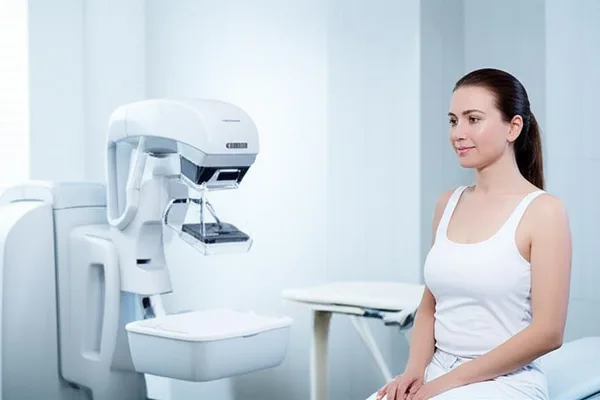
2. Then you will be taken to the room where the ``Gamma Camera'' is located. There you will be asked to sit on a chair in front of the testing device.
3. Next, a medical staff member will insert a small IV line into a vein in your arm and inject the radioactive tracer through it. This will feel like a small ant bite, but it won't be very painful.
4. You will need to wait about three to five minutes for the `(Tracer)` to travel to your breast tissue.
5. Then the scan begins. This can take about 20 to 40 minutes. During the scan, the device will gently press on your breast. But this will not hurt. There will be no hard pressing here like in a mammogram. So don't worry about it.
What happens after the MBI?
After the MBI scan is complete, the IV line in your arm will be removed. You can then put your clothes back on. Your medical team will advise you on what to do next. You may need to see your doctor again for a follow-up visit, or you may be referred to another specialist.
Are there any risks in MBIs? Let's find out a few things.
During an MBI scan, you are exposed to a small amount of radiation because of the tracer. Continued exposure to radiation can increase the risk of cancer.
However, the radiation exposure during MBI is very low and is considered safe. In fact, it is about the same as the radiation exposure during a mammogram. In most cases, the benefits of detecting breast cancer early far outweigh the risks of this small amount of radiation exposure.
A very small number of people may have an allergic reaction to the tracer. If this happens, they may show symptoms such as:
- Skin conditions such as rashes, itching, or eczema.
- Nausea or vomiting.
- Breathing difficulties or heart rhythm irregularities (these are very rare, occurring in a small percentage of people, around 0.01% - 0.04%).
If something like this happens, you should immediately inform the medical staff.
What are the results of MBI?
A radiologist will carefully examine your MRI images to see if there are any cancer cells. If any areas on the images are "lighted up," your doctor may take a small sample of tissue (a biopsy) from those areas and examine it. This biopsy will confirm whether the cells are actually cancer cells. The results of the biopsy will also help determine the best treatment for you.
If there are no abnormalities on your MBI scan, it is usually considered a "normal" result. You can then talk to your doctor about your breast cancer screening schedule, such as when you should have your next mammogram.
How accurate is MBI?
Studies have shown that MBI can detect about 90% of breast cancers in patients with suspected breast cancer. It can detect 82% of very small cancers, less than 10 millimeters in diameter. Studies have shown that these results are as accurate as a breast MRI. Studies have also shown that MBI can detect two to three times more breast cancers than a mammogram alone.
How long does it take to get results?
You can ask your doctor how long it will take to get the results, and how they will let you know. In most cases, you will get the results within a week of the test.
Finally, things to remember
In general, getting regular mammograms is the best way to detect breast cancer early. However, sometimes, when a mammogram alone is not enough, a test called Molecular Breast Imaging (MBI) can be very useful to get additional information.
Remember, there are other breast imaging tests, such as breast ultrasound and breast MRI, that can provide detailed information about abnormalities in the breast. Each test has its pros and cons. Therefore, discuss all of these with your doctor. Together, you can choose the test methods that are right for you, and prevent breast cancer from developing, or if it does develop, detect it early and treat it successfully. Early detection is the most important thing! Don't be afraid, and if you have any doubts, seek medical advice.
` Breast cancer, MBI, molecular breast imaging, mammogram, cancer screening, women's health, nuclear medicine




💬 Comments (0)
No comments yet. Be the first to share your thoughts here.
Add Your Comment