
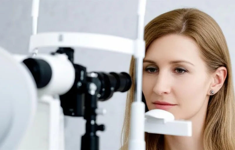
Have you ever had an eye exam? When you do, your doctor may tell you to do a special test. One such special, very important test that we're going to talk about today is called the OCT. Don't worry, it's very simple and painless.
What is this (OCT)?
Simply put, Optical Coherence Tomography (OCT) is a special method that can take pictures of what's inside your eye. It's like doing an ultrasound scan to look inside your body, but this one is done on your eye. It uses infrared light, which is harmless to the eye and invisible to us, to take pictures of the layers inside your eye. It can especially clearly see the very important parts at the back of the eye, such as the retina and optic nerve . It's like a real eye scan. With this, doctors can measure the depth of your eye, examine various parts, and diagnose and treat any medical conditions.
This is sometimes called "ocular coherence tomography", "ocular" meaning related to the eye. Nowadays, OCT technology is used not only for eyes, but also for other diseases. For example, it is used for things like blood vessel examination (Angiography). Specialists in cardiology, neurology, and oncology have also recognized the benefits of this method.
When should I have this (OCT) test?
Your doctor may suggest an OCT test based on your symptoms or something they see during an eye exam. For example, if you have a change in your vision or if your doctor suspects there is something wrong with your eye, they can look at what is going on.
Also, there are some age-related eye problems that come with age, so for those who are at risk for them, or who are at risk for other eye diseases, the doctor may recommend that they have an OCT scan along with their regular eye exam. Then they can take these scans from time to time and compare them to see if there are any changes. It's like keeping a record of the health of our eyes.
What types of eye diseases can OCT help diagnose and treat?
Ophthalmologists use this (OCT) test to examine the retina and optic nerve at the back of your eye. It can help diagnose a variety of conditions that can affect your vision. Here are a few examples:
- Glaucoma: This is a condition where the pressure inside the eye increases and can damage the optic nerve. OCT can be used to see the extent of this damage and the thickness of the nerve fiber layer.
- Macular Degeneration: This is a common condition that occurs as we age. The macula, the part of the eye where we see clearly, is damaged. OCT can help determine the type, whether it is dry or wet, and the extent of the damage.
- Diabetes-related macular edema: A major complication that can occur in people with diabetes. The macula becomes swollen due to fluid accumulation. Optical coherence tomography (OCT) can accurately measure the extent of this swelling.
- Diabetes-related Retinopathy: Diabetes can damage the blood vessels in the retina and cause new blood vessels to form. This damage and fluid buildup can be clearly seen with (OCT).
- Eye Cancer: This also helps to identify some types of eye cancer and see their extent.
- Optic Atrophy: This condition can occur due to damage to the optic nerve due to various reasons. Optical coherence tomography (OCT) can show the thinning of the nerve fibers.
- Retinal Detachment and Retinal Tears: This is very important to identify things like tears and small tears before the retina detaches.
- Posterior Vitreous Detachment: This is also common with age. Sometimes an OCT scan can help to see if it is affecting the retina.
- Central Serous Retinopathy: This is a condition where fluid builds up under the retina, which can cause blurred vision. This fluid layer can be clearly seen with (OCT).
- Cystoid Macular Edema: The formation of small, fluid-filled cysts in the macula. This can occur for a variety of reasons.
- Bull's Eye Maculopathy: A condition that causes damage to a specific "target" (bull's eye) pattern in the macula. This can be caused by certain medications.
- Retinoschisis: A separation of the layers of the retina from each other.
Sometimes, doctors also use OCT to look at the front of the eye, such as the cornea and iris. This can help identify defects in the front of the eye or help plan eye surgery.
How does this (OCT) test work?
It's very simple. Have you heard of an ultrasound scan? It uses sound waves to send into the body and create a picture as they bounce back. OCT is similar, but instead of using sound waves, it uses invisible red light . When these light rays bounce back and hit the tissues inside your eye, they create a 3D cross-sectional image. This means you can see what's inside your eye layer by layer . Think of it like cutting a cake in half.
This does not harm the eyes in any way. Nothing gets into the eyes. It is a very safe method.
What happens during an (OCT) test?
This is a very quick test that can be completed in just a few minutes. It is done right in your doctor's office. No special preparation is required. It can even be done right after your regular eye exam.
Before the test, you may be given eye drops to dilate your pupil. This will help you see the inside of your eye more clearly. You will then be asked to sit in front of the scanner and rest your chin on a support attached to it. You will be asked to stare straight at a green light.
The (OCT) scanner scans one eye at a time. You will see a red line during the scan. You will not feel anything, and nothing will hit your eye. It takes only a minute or two to scan one eye. It is important to keep your eye in one place without moving your head during this time. This is the only way to get a clear picture.
Are there any side effects to this?
No, the (OCT) test itself has no risks or side effects. It is 100% safe. However, as I mentioned earlier, if you use medication to enlarge the pupil of the eye (pupil dilation), you may experience some minor discomfort for a while afterward. For example:
- Sensitive to light: This means that your eyes may feel blue when you go out in the sun. Wearing sunglasses can help reduce this.
- Blurry vision: This is especially noticeable when looking up close.
These usually resolve completely within a few hours (about 4-6 hours). Some people may also experience a headache.
So, if you are being tested with such medication, it is best to have someone else drive you home, or avoid driving, because it takes a while for your vision to return to normal.
What kind of results are obtained? What do they tell us?
Your eye specialist (OCT) will carefully study the images taken from the scan. The images are like a map of the inside of your eye. If you have had a previous scan, they will compare these new images with those images to see if there has been any change, improvement, or progression of the disease. They will then talk to you as soon as possible to explain this.
These results can help your doctor determine if you have a condition affecting your retina or optic nerve, and if so, how far it has spread and how serious it is. Using this information, your doctor will plan your next course of treatment. They may recommend starting medication, laser treatment, or eye injections.
When should I see my doctor?
It is very important to have your eyes checked regularly. Visit your doctor at regular intervals (every 6 months, once a year, for example). This is when your doctor can check for any changes in your vision. If you have any unusual symptoms or notice any changes in your vision, you should definitely tell your doctor. Don't ignore it, even if it's a small thing.
If you have any of the symptoms below, don't panic and see a doctor immediately:
- New or persistent eye pain.
- If you have difficulty seeing clearly, if the way colors appear has changed, or if things appear blurry.
- If you frequently see things that look like floaters before your eyes, or if you see flashes of light.
- If you suddenly lose your vision, or if part of your vision seems to be black.
So, the final take-home message:
Simply put, this (OCT) test is a very quick, easy, painless, and highly advanced test. It takes layer-by-layer, three-dimensional (3D) images of the inside of your eye, especially the sensitive parts at the back of the eye, such as the retina and optic nerve, and allows you to see them as finely as a thousandth of a millimeter.
This allows doctors to take measurements inside the eye, see damage to the optic nerve, and detect conditions that may be affecting your vision at an early stage and plan treatment. So, if a doctor asks you to have this test, there's no reason to be afraid. It can be a very important step in the health of your eyes and in preserving your precious vision.
Take care of your eyes! Don't forget to keep getting your eyes checked.
` Eye exam, OCT test, retina, glaucoma, diabetic eyes, eye health, scan test


💬 Comments (0)
No comments yet. Be the first to share your thoughts here.
Add Your Comment