

Have you ever heard of fluid filling up around the heart? It sounds a little scary, doesn't it? This condition can actually be very dangerous at times. But don't worry, there is a special medical treatment that can even save a life in such cases. That's the treatment we're going to talk about today.
What is Pericardiocentesis? Let's understand it simply
Pericardiocentesis is a procedure that removes excess fluid from the sac around your heart (called the pericardium in medical terms). This is a very important procedure, as this type of fluid can sometimes cause the heart to stop.
Imagine that our heart is inside a protective sac. There is normally a small amount of fluid inside this sac to help the heart move and beat. That is not a problem. But if for some reason this amount of fluid increases too much, we call it Pericardial Effusion .
This increase in water is like putting your heart inside a water balloon and filling the balloon with more and more water. Gradually, the heart loses its ability to beat, and the heart becomes congested.
This blockage is the most dangerous. We call this Cardiac Tamponade . This is a medical emergency. Because it can cause the heart to stop working, and death can occur within a few hours or even minutes.
So, in the Pericardiocentesis treatment, a thin needle is inserted through the chest, into the sac around the heart, and the excess fluid is removed. Sometimes a tube may be placed to remove some of the fluid.
Who needs this treatment? What are the reasons?
There can be a number of reasons why fluid can build up around the heart in this way. Here are some of the main reasons why this treatment may be necessary.
| Reason | Description |
|---|---|
| Cancer status | Especially severe cancers and heart cancer. |
| Heart attack | In cases like when a wall of the heart bursts due to a heart attack. |
| Infections | Infection of the sac around the heart due to tuberculosis, bacterial or viral infections (such as COVID-19, HIV). |
| Immune system diseases | Diseases such as Lupus and Rheumatoid Arthritis. |
| Other medical conditions | Heart or kidney failure, hypothyroidism. |
| Other reasons | Some medications, radiation treatments, or sometimes no cause can be found. |
What happens before treatment?
Depending on whether this is an emergency or not, the preparation process may vary slightly. If it is not an emergency, your doctor will give you a day and time. Most often, you will be asked to stop eating and drinking 8 hours before the procedure.
Let's look at some of the things that are usually done before treatment.
- Inserting an IV line: A small tube (IV line) is inserted into your vein so that things like saline or medications can be given.
- Skin preparation: The area where the needle will be inserted is shaved and the area is thoroughly cleaned with an antiseptic solution. This is done to prevent infection.
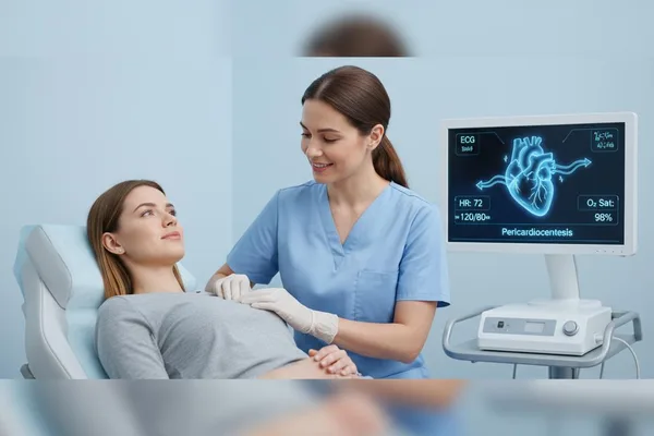
- Vital signs monitoring: Sensors will be attached to monitor your blood pressure, breathing, heart rate, and oxygen levels . You will also be connected to an ECG (electrocardiogram) to monitor the electrical activity of your heart.
- Oxygen administration: If necessary, oxygen is administered through a small tube placed in the nose.
What happens during treatment?
This treatment is performed by a team of doctors, nurses, and other technicians.
Using imaging technology to help
Before inserting the stent, the doctor uses imaging technology to find the exact location of the fluid around the heart. This is often done using an ultrasound (echocardiogram) . This helps determine the safest and easiest place to insert the stent. This greatly reduces the risk of damage to the heart or other organs.
How to insert the needle
Unless it's an emergency, a local anesthetic will be injected into the area where the needle is inserted. So you won't feel any pain. You may feel a little pressure.
There are several places where the needle can be inserted. The most common place is under the middle bone of the chest (sternum). From there, it can easily enter the sac around the heart.
Once the needle is properly inserted into the sac, the doctor uses a syringe to slowly draw out the fluid. This usually takes about 10-20 minutes. If there is a lot of fluid, a small tube (catheter) may be inserted to drain it for a day or two.
After the work is done, the pin or tube is removed and a small piece of plaster is applied to cover the area.
What happens after treatment?
The medical team will take good care of you after treatment.
- Fluid testing: The extracted fluid sample is sent to the laboratory to determine the cause of the fluid buildup.
- Re-scan: A repeat ultrasound (echocardiogram) scan will be done to see if all the fluid has been removed.
- Recovery: You will be told to rest for about 12-24 hours. You will start to feel much better soon after the fluid is removed.
What are the benefits and risks of this treatment?
Benefits
- Can save lives: This is a life-saving treatment in an emergency like Cardiac Tamponade.
- The cause can be found: By examining the fluid, the underlying cause of the illness can be found. This is very important for future treatment.
- Quick and simple: This is quicker than major surgery. Since there is no major incision, the recovery time is shorter.
Risks
As with any medical procedure, there are some risks involved. But remember, these risks are very low because technology like ultrasound is used.
- Damage to nearby organs such as the heart, lungs, or liver from the shell (this is very rare).
- Arrhythmia occurs when the heart's electrical system is affected.
- Infection at the site of insertion of the needle.
- Damage to a major blood vessel.
But compared to the potential harm of not having this treatment, these risks are relatively small. Your doctor will weigh all of these factors and decide what is best for you.
When to seek medical advice again
If you experience any symptoms after returning home after treatment, you should immediately go to the hospital's Emergency Treatment Unit (ETU) .
| Symptoms that require immediate attention | |
|---|---|
| Symptoms of Cardiac Tamponade | |
| |
| Signs of an infection (Sepsis) | |
|
Is this treatment painful?
You won't feel any pain because the needle is injected with a numbing medicine. You may feel a slight pressure when the needle goes in, but it's not painful.
Take-Home Message
- Pericardiocentesis is a sometimes life-saving treatment that removes unwanted fluid that accumulates around the heart.
- This accumulation of fluid (pericardial effusion) can cause cardiac tamponade, an emergency.
- This treatment is very safe because it is performed using technology like ultrasound .
- If you develop symptoms such as chest pain, difficulty breathing, or fever after treatment , see a doctor immediately or go to the hospital's Emergency Treatment Unit (ETU).
- If you have any questions about this treatment, don't hesitate to ask your doctor.




💬 Comments (0)
No comments yet. Be the first to share your thoughts here.
Add Your Comment