

Most of us have had back pain or neck pain at some point in our lives, right? Sometimes your doctor may ask you to take an X-ray of your back to find out exactly what is causing the pain. So when you hear about this X-ray, some people are a little scared. "What is this looking at? What should I know before doing this? Is there any danger?" Many questions come to mind. Let's talk about all of this very simply and in a friendly way today.
What exactly is a back X-ray?
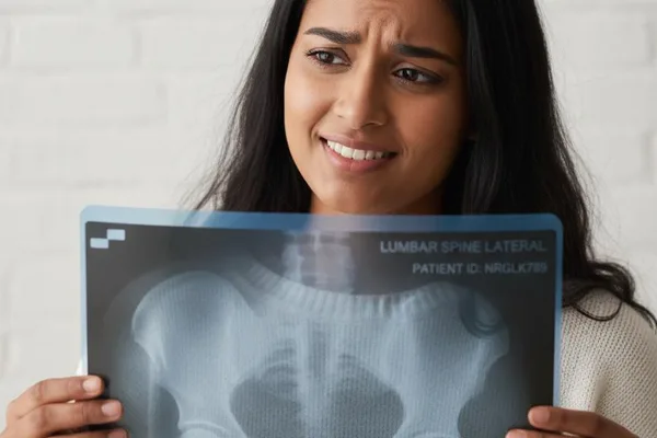
Simply put, a spine X-ray is a test that takes pictures of the bones in your neck and back, called vertebrae. It uses a special type of electromagnetic waves (X-rays).
Think of it like taking a picture of your spine. Your bones, called vertebrae, absorb these X-rays, so they appear white in the picture. But your soft tissues, like muscles and fat, appear gray because the X-rays pass through them more easily. By looking at this picture, your doctor can get a good idea of what is causing your neck or back pain.
Different names for the parts of our spine and X-rays
The name of the X-ray taken will vary depending on where your back pain is. Our spine is not one long bone, it is a collection of several parts. Let's see what those parts are and what the names of the X-rays taken related to them are.
| Spine Section | Location and simple description | Name of the relevant X-ray |
|---|---|---|
| Cervical spine | Your neck is made up of 7 vertebrae. | Cervical spine X-ray |
| Thoracic spine | The 12 vertebrae in your upper and middle back. | Thoracic spine X-ray |
| Lumbar spine | The 5 vertebrae in the lower back (lumbar) area. | Lumbosacral spine X-ray |
| Sacral spine/Sacrum | Five small vertebrae fused together, located just below the lower back. | Often covered by the Lumbosacral X-ray itself. |
| Coccyx/tailbone | The small bone at the lowest end of the spine. | Coccyx X-ray |
So you understand, the part that corresponds to where your pain is is what the doctor refers you to for an X-ray.
What diseases can be detected with this X-ray?
A back X-ray is not just a picture. It can provide a doctor with a lot of important information.
An X-ray can provide a clear picture of the condition of your spine's bones. This can be a great help in identifying the exact cause of your pain.
Let's see what are the main medical conditions that can be diagnosed with this X-ray.
| Medical condition | A simple explanation |
|---|---|
| Fractured spine | A fracture or split in a vertebra due to a fall or accident. |
| Osteoporosis | Bone density decreases with age or due to other reasons, making them more likely to break easily. |
| Arthritis | Pain caused by inflammation or swelling of the joints between the vertebrae. |
| Spinal tumors | Abnormal cell growths in the spine. |
| Slipped disk | A herniation of the cushion-like part (disc) between two vertebrae. |
| Spinal curvature disorders | The natural position of the spine changes and becomes curved forward (Lordosis), backward (Kyphosis), or sideways (Scoliosis). |
| Congenital disorders | Certain problems with the spine that occur at birth, for example `(Spina bifida)`. |
Do I need to prepare before the X-ray?
In most cases, no special preparation is needed for a spine X-ray. You can eat and drink normally. However, if your doctor gives you any special instructions, you should follow them.
But there are a few things to keep in mind:
- Clothing: Wear as light and comfortable clothing as possible.
- Jewelry: It's easy to leave metal jewelry like necklaces and earrings behind when you leave home. You'll be asked to remove them at the X-ray facility anyway. Metal items can interfere with the X-ray image.
- You may be asked to take off your bed clothes and put on a special gown that they provide.
What happens when an X-ray is taken?
This is a very simple and quick process, usually taking about 15 minutes.
1. First, the radiographer/radiologist will explain what needs to be done to you.
2. You will be asked to remove any metal objects (such as jewelry, belts, coins) from your body.
3. You will then be positioned for the X-ray. Most of the time, you will be asked to lie down on a table. Sometimes, you may be asked to stand.
4. To protect the parts of the body that do not need to be X-rayed, a lead apron is placed over them. This prevents unnecessary radiation from entering the body.
5. The radiologist will go behind a protective screen and give you instructions. For example, they may say something like, "Hold your breath for a moment." This will help you to take a clear picture without moving.
6. You won't feel any pain when you get the X-ray. However, if you already have back pain, you may feel a little uncomfortable when they position you. But they will try to make you as comfortable as possible.
Is it risky to get a back X-ray?
This is a problem that many people have. It's normal to be afraid of radiation when you hear the word X-ray.
But the truth is, a medical X-ray emits a very small amount of radiation . This amount is usually not enough to harm the body. You also get extra protection from the lead apron that you wear.
However, if you have any doubts about this, do the following:
- Talk to your doctor about this openly.
- Keep a record of any X-rays you have taken previously.
- Most importantly: If you are pregnant, or have any suspicion that you may be pregnant, be sure to tell your doctor and radiologist before having the X-ray. This is very important.
When will I receive the report?
After the X-ray images are taken, a radiologist will examine the images and prepare a report. This report and the images will be sent to your treating doctor. This can usually take a day or two.
Your doctor will review the report and explain to you clearly what is in it, what is causing your pain, and what treatment is needed next. If more information is needed, your doctor may also recommend other tests, such as a ``Myelogram'' or ``CT scan''.
Back pain and neck pain can be a lifelong problem. So this X-ray is a very important test that will help your doctor find the exact cause of this pain at a level that is not visible to the naked eye. So don't be afraid of this, and do this test as your doctor says.
Take-Home Message
- A back X-ray is a simple, painless test that takes pictures of the bones (vertebrae) in your back and neck.
- This can detect many things, such as spinal fractures, bone thinning, joint pain, and spinal strains.
- There is no special preparation required before going for an X-ray, but you will need to remove any metal objects such as jewelry.
- If you are pregnant or may become pregnant, be sure to inform the doctor and radiologist.
- The amount of radiation from this test is very low, so there is usually nothing to worry about.
- Your doctor will explain to you the full details of your X-ray report and what to do next.




💬 Comments (0)
No comments yet. Be the first to share your thoughts here.
Add Your Comment