

Consider the subtle changes you notice in the mirror every day: a dry patch on your elbow during dry winter months, a brittle fingernail that splits after washing dishes, or goosebumps rising across your forearms during a sudden cold breeze. While our society often frames skin, hair, and nails as mere cosmetic features or aesthetic goals, medical professionals recognize them as something vastly more consequential. Together, they constitute the integumentary system—the human body’s largest, heaviest, and most interactive organ system. Weighing about 3.6 to 4.5 kilograms (8 to 10 pounds) and covering nearly 1.5 to 2.0 square meters of surface area in the average adult, this dynamic biological barrier forms your frontline defense against microbial invasion, fatal fluid depletion, ultraviolet radiation, and thermal collapse. Understanding the physiology of this protective fortress is essential to protecting your overall physical health.
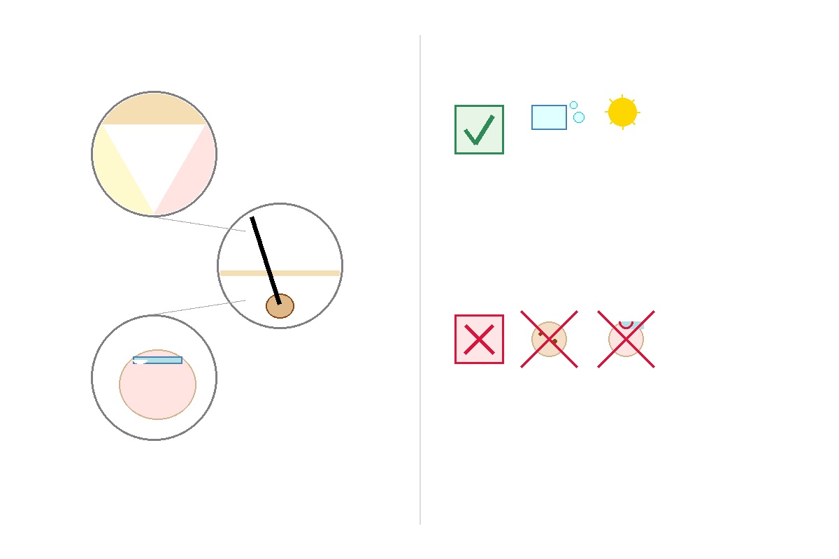
Key Clinical Takeaways.
- Primary Barrier Function: The stratified layers of the epidermis—anchored by keratin-packed corneocytes and intercellular lipids—form an impermeable seal preventing transepidermal water loss (TEWL) and blocking microbial penetration.
- Endocrine & Metabolic Synthesis: Ultraviolet B (UVB) radiation initiates the conversion of cutaneous 7-dehydrocholesterol into cholecalciferol (pre-vitamin D3), essential for systemic calcium absorption and skeletal homeostasis.
- Diagnostic Window: Pathologic alterations in nails (e.g., clubbing, splinter hemorrhages) and skin (e.g., acanthosis nigricans, erythema multiforme) often provide the earliest clinical signs of severe internal cardiopulmonary, metabolic, or autoimmune diseases.
- Thermoregulatory Precision: Through an elaborate network of eccrine sweat glands and dermal microvascular vasoconstriction/vasodilation, the integumentary system maintains core internal body temperature within narrow physiologic boundaries (36.5°C to 37.5°C or 97.7°F to 99.5°F).
Know your body mass index (BMI) and weight classification
What Is the Integumentary System? Structural Anatomy and Tissue Layers.
The integumentary system comprises the cutaneous membrane (the skin) and its specialized accessory appendages: hair follicles, sebaceous glands, sudoriferous (sweat) glands, and nails. Histologically, the skin consists of three primary architectural tiers, each uniquely engineered to perform distinct physiological duties:
1. The Epidermis: The Waterproof Cellular Shield.
The outermost layer is an avascular, stratified squamous epithelium dominated by keratinocytes (>90% of cells), which originate in the deepest basal layer and migrate outward over about 28 to 40 days, progressively flattening, losing their nuclei, and synthesizing dense keratin before desquamating. It is subdivided into five distinct strata in thick skin (palms and soles) and four in thin skin:
- Stratum Basale: A single layer of actively dividing stem cells anchored to the basement membrane, interspaced with pigment-producing melanocytes (synthesizing photoprotective melanin) and mechanoreceptive Merkel cells for fine touch.
- Stratum Spinosum: Several rows of polyhedral keratinocytes joined by robust intercellular bridges (desmosomes), interspersed with immunocompetent Langerhans cells (epidermal dendritic cells that phagocytose antigens and activate T-cells).
- Stratum Granulosum: Keratinocytes accumulating dense keratohyalin granules and lamellar bodies, extruding lipid-rich compounds that establish the critical hydrophobic extracellular permeability seal.
- Stratum Lucidum: A thin, translucent, clear band present only in the thick friction surfaces of the fingertips, palms, and foot soles.
- Stratum Corneum: The outermost mechanical shield made of 15 to 30 layers of dead, flattened, anucleated corneocytes embedded within a specialized lipid mortar (ceramides, cholesterol, and free fatty acids)—the classic "brick-and-mortar" structure.
2. The Dermis: The Structural and Vascular Scaffold.
Positioned directly beneath the epidermal basement membrane, the dermis is a durable, vascularized fibroelastic tissue network primarily produced by fibroblasts:
- Papillary Dermis: The superficial zone consisting of loose areolar connective tissue with finger-like dermal papillae projecting toward the epidermis. It houses extensive capillary loops delivering oxygen and nutrients to the avascular epidermis, alongside Meissner’s corpuscles (rapidly adapting touch sensors).
- Reticular Dermis: The thicker, deeper layer composed of dense irregular connective tissue. Rich in Type I and Type III collagen bundles, woven elastin fibers, and proteoglycan ground substance, it delivers tensile strength, structural resilience, and elasticity while housing hair bulbs, nerve fibers, Pacinian corpuscles (pressure and vibration receptors), and sweat/sebaceous glands.
3. The Hypodermis (Subcutis): Cushioning and Energy Reserve.
Although technically classified as a subcutaneous tissue layer beneath the true skin, the hypodermis is clinically inseparable from integumentary performance. Dominated by lobules of mature adipocytes separated by fibrous septa, it anchors the skin to underlying muscle fascia and periosteum, stores surplus metabolic energy, provides physical shock absorption over bony prominences, and serves as an effective thermal insulator against ambient cold.
Accessory Appendages: The Functional Roles of Hair, Glands, and Nails.
Far from ornamental extensions, cutaneous appendages are specialized epithelial down-growths that carry out essential physiological tasks:
Hair Follicles and Hair Fibers.
Hair consists of dead, filamentous columns of tightly compacted, heavily keratinized epithelial cells divided into a cuticle, cortex, and central medulla. Hair undergoes cyclical phases: anagen (growth phase, lasting 2 to 7 years), catagen (regression phase, 2 to 3 weeks), and telogen (quiescent resting phase, 2 to 4 months):
- Cranial Thermal Protection: Scalp hair shields the calvarium from direct ultraviolet solar radiation and prevents radiant heat loss.
- Vibrissae (Nasal and Aural Hair): Specialized coarse hairs lining the anterior nares filter out coarse airborne particulates, spores, and irritants before air enters the lower respiratory tree.
- Sensory Perception: Sensory nerve fiber plexuses wrapped around the hair follicle bulb (peritrichial nerve networks) detect microscopic mechanical displacement of the hair shaft, alerting the central nervous system to tactile contact or parasites on the skin surface.
- Arrector Pili Action: Tiny smooth muscle bands connected to each hair follicle contract during sympathetic nervous system arousal or cold exposure, producing cutis anserina ("goosebumps") to trap an insulative boundary layer of still air against the skin surface.
Cutaneous Glands.
- Sebaceous Glands: Holocrine glands that secrete an oily, lipid-rich substance (sebum) directly into hair follicles. Sebum acts as an endogenous emollient, lubricating hair and skin while imparting weak antibacterial and antifungal properties through antimicrobial peptides and free fatty acids.
- Eccrine Sweat Glands: Simple coiled tubular glands distributed over nearly the entire cutaneous envelope (highest density on palms, soles, and forehead). They secrete hypotonic fluid (composed of 99% water, sodium, chloride, potassium, urea, and antimicrobial dermcidin) for rapid evaporative thermoregulation.
- Apocrine Sweat Glands: Larger glands located primarily in the axillary and anogenital zones that secrete viscous, odorless fluid containing lipids and proteins. These secretions produce typical body odor when cleaved by cutaneous commensal bacteria.
The Nail Apparatus.
The human nail is a plates of hard alpha-keratin that rests upon the richly innervated and vascularized nail bed. The germinal matrix situated beneath the proximal nail fold drives cellular proliferation and continuous nail plate growth (fingernails grow roughly 3 mm per month. Toenails grow at roughly one-third that rate). Nails provide rigid mechanical counter-pressure that enhances fine tactile sensitivity in the distal digital pulp, facilitates precise manipulation of small objects, and shields digital phalanx bones against blunt trauma.
Integumentary Cross-Talk: How Skin Supports Other Organ Systems.
The skin does not work in isolation. It maintains direct functional links with nearly every internal physiological system:
- The Immune System (Cutaneous Immunity): The skin is a major immunological organ housing Skin-Associated Lymphoid Tissue (SALT). Epidermal Langerhans cells capture external microbial antigens and migrate to regional draining lymph nodes to prime naive T-lymphocytes. Meanwhile, keratinocytes constitutively express pattern-recognition receptors (e.g., Toll-like receptors) and secrete antimicrobial peptides (defensins, cathelicidins) that neutralize bacteria upon contact.
- The Endocrine & Skeletal Axis (Vitamin D Synthesis): When sunlight hits bare skin, solar UVB radiation (wavelengths 290 to 315 nm) breaks the B-ring of cutaneous 7-dehydrocholesterol to form pre-vitamin D3, which thermally isomerizes into cholecalciferol (vitamin D3). This molecule is then hydroxylated in the liver to 25-hydroxyvitamin D [25(OH)D], and finally converted by the kidneys into calcitriol [1,25(OH)2D], the active hormone that enables intestinal absorption of dietary calcium and phosphorus to prevent rickets in children and osteomalacia/osteoporosis in adults.
- The Nervous System (Somatosensory Input): Operating as our largest sensory surface, the skin is packed with specialized mechanoreceptors, thermoreceptors (transient receptor potential ion channels), and nociceptors (free nerve endings conveying acute and slow pain). This constant sensory stream provides the brain with continuous proprioceptive feedback and triggers rapid defensive withdrawal reflexes when exposed to noxious stimuli.
Emergency Red Flags: When to Seek Immediate Care.
Certain cutaneous symptoms indicate critical, life-threatening systemic emergencies. Call 911 (US/Canada), 999 (UK), or proceed directly to your nearest Emergency Department (ER / A&E) if you experience:
- Suspected Necrotizing Fasciitis: Rapidly advancing erythema (redness) and severe pain that is grossly disproportionate to physical examination findings, accompanied by skin crepitus (crackling under pressure), purplish bullae, cutaneous anesthesia, or systemic collapse.
- Severe Cutaneous Adverse Reactions (SCARs): Extensive sloughing or blistering of the skin (positive Nikolsky sign), painful mucosal erosions involving the mouth, eyes, or genitals, high fever (>38.5°C / 101.3°F), and skin tenderness suggestive of Stevens-Johnson Syndrome (SJS) or Toxic Epidermal Necrolysis (TEN).
- Anaphylactic Integumentary Signs: Rapidly evolving urticaria (hives), severe facial, lip, or tongue edema (angioedema) accompanied by stridor, wheezing, respiratory distress, throat tightness, or dizziness.
- Meningococcal Purpura Fulminans: Non-blanching petechial or purpuric rash (spots that do not turn pale when pressed firmly with a clear glass) alongside high fever, neck stiffness, photophobia, and confusion.
- Extensive Thermal Burns: Second- or third-degree burns involving greater than 10% of total body surface area (TBSA), or burns involving the face, hands, feet, perineum, or major joints.
Evidence-Based Guidelines: Optimal Daily Care for Skin, Hair, and Nails.
Maintaining the integrity of the integumentary barrier requires evidence-based personal care practices that support natural barrier dynamics without disrupting cutaneous physiology.
| Area of Care. | Clinical Best Practice (Do). / Harmful Habit or Pitfall (Don't). |
|---|---|
| Skin Cleansing & Hydration. | Clinical Best Practice (Do).: Wash with lukewarm water and soap-free, pH-balanced syndet cleansers (pH 5.5). Apply lipid-rich emollient moisturizers (containing ceramides, hyaluronic acid, or petrolatum) within 3 minutes of bathing to seal in epidermal hydration. Harmful Habit or Pitfall (Don't).: Do not bathe with prolonged hot water or use alkaline, fragrance-heavy bar soaps (pH > 9.0), which strip stratum corneum intercellular lipids and elevate the skin's acidic mantle, promoting bacterial overgrowth. |
| Photoprotection. | Clinical Best Practice (Do).: Apply broad-spectrum sunscreen (protecting against both UVA and UVB) with an SPF ≥ 30 daily to all sun-exposed skin, reapplying every 2 hours during outdoor activity. Wear wide-brimmed hats and UPF-rated clothing. Harmful Habit or Pitfall (Don't).: Never use commercial indoor tanning beds; the concentrated UVA exposure increases your lifetime risk of melanoma by up to 75% when initiated before age 35 and rapidly degrades dermal collagen, causing premature solar elastosis. |
| Nail Hygiene. | Clinical Best Practice (Do).: Trim fingernails and toenails straight across with dedicated clippers, slightly rounding the edges with an emery board to prevent ingrown nails (onychocryptosis). Keep subungual areas clean using a soft nail brush and water. Harmful Habit or Pitfall (Don't).: Avoid habitual nail biting (onychophagia), picking at hangnails, or aggressively pushing back or trimming the cuticle (eponychium). The cuticle forms a biological seal that protects the nail matrix against candidal and bacterial paronychia. |
| Hair & Scalp Maintenance. | Clinical Best Practice (Do).: Wash the scalp based on person sebum production rates. Apply gentle conditioners to the hair shafts to reduce inter-fiber friction. Use wide-toothed combs on wet hair to minimize mechanical tensile fracture. Harmful Habit or Pitfall (Don't).: Do not pull hair into excessively tight ponytails, tight braids, or extensions that apply prolonged tension to hair follicles, which causes irreversible traction alopecia. Avoid plucking nasal hairs directly with tweezers, which can cause nasal vestibulitis. |
| Lesion Surveillance. | Clinical Best Practice (Do).: Conduct monthly full-body skin self-exams using the clinical ABCDE rule for pigmented lesions: Asymmetry, Border irregularity, Color variation, Diameter > 6 mm, and Evolving size, shape, or symptoms. Harmful Habit or Pitfall (Don't).: Do not scratch, pick, or attempt self-excision of suspicious cutaneous moles, persistent non-healing ulcers, or recurring nodules. Consult a certified dermatologist for professional dermoscopy and biopsy. |
Authoritative References & Further Reading.
- National Health Service (NHS). Nail problems and common conditions. NHS UK, 2024. Available from:
- National Institutes of Health (NIH) / MedlinePlus. Skin Conditions and Cutaneous Anatomy. National Library of Medicine, 2023. Available from:
- Cleveland Clinic Staff. Integumentary System: Parts, Function & Conditions. Cleveland Clinic Medical Center, 2023. Available from:
- Bolognia JL, Schaffer JV, Cerroni L. Dermatology, 4th Edition. Elsevier Health Sciences, Philadelphia, 2018.






No comments yet. Be the first to share your question or experience below.