A special camera that looks inside the body: Everything about Nuclear Medicine Imaging!

A special camera that looks inside the body: Everything about Nuclear Medicine Imaging!

Have you ever heard of a test called 'Nuclear Medicine Imaging'? You probably heard about it when your doctor told you to do such a test. It really sounds a little scary when you hear this name, doesn't it? 'Nuclear' when you say it like that. But don't worry, today we will talk about it in detail, in Sinhala that you can understand, and very simply. Just like talking to a friend.

What is this so-called Nuclear Medicine Imaging?

Simply put, this is a special method used to look at the organs inside our bodies. But unlike other scans, this one has a slight difference. Here's what happens: First, you are given a very small amount of a 'radioactive tracer'. This is most often given through an injection into a vein. Sometimes it is given as a drink, depending on the test being performed. This is not a medicine or a liquid that dyes the body, is it? When this is given, it travels through our blood, goes to the organ or tissue that we want to examine, and is deposited there.

Then, you are placed under a special camera called a 'gamma camera', which detects the subtle radiation emitted by the substance that was inserted into your body. This camera is very sensitive. That radiation information is sent to a computer and turned into beautiful images. It's like a 'map' of an organ inside our body. It's by looking at those images that doctors can find out if there is any problem in your body or if an organ is working properly.

Now you might be thinking, 'Oh, radioactive, it's scary, it must be bad for your body.' But the truth is, the amount of radiation in the material used for this test is very low . That is, it is the same amount of radiation that we are usually exposed to in our daily environment, and sometimes even less. Also, these materials usually do not cause any side effects. These materials are eliminated from the body after a while. So there is no reason to be unnecessarily afraid.

Why do we need this type of test (Nuclear Imaging Test)?

Well, this test is mainly used to diagnose various diseases. Sometimes it is also used to treat and see how effective the current treatment is. Just imagine how much this test can help our doctors find. Here are some of the diseases and conditions that this test can detect:

  • Some blood diseases (e.g., some types of anemia, blood clotting problems).
  • Thyroid diseases, especially conditions such as hypothyroidism, where the thyroid gland does not function properly.
  • Heart disease (e.g., reduced blood flow to the heart muscle, damage to the heart after a heart attack).
  • Gallbladder diseases (e.g., cholecystitis, bile duct obstruction).
  • Lung problems (e.g., blood clots in the lungs, decreased lung function).
  • Bone-related problems, such as bone infections, hard-to-find fractures, and whether cancer cells have spread to the bones.
  • Kidney diseases, for example, kidney infections, scarring, and urinary tract obstruction.
  • Identify different types of cancer and see how far the cancer has spread (staging).

In addition to this, as I mentioned earlier, this ``Nuclear Medicine Imaging'' technology is also used for treatment methods. An example is the ``Radioimmunotherapy'' treatment method. In this, radiation and immunotherapy are combined, and radiation is sent to a targeted area, just like cancer cells, for treatment. This means that damage to healthy cells can be minimized.

What is the difference between this and other radiologic tests?

This is a very important question for you to understand. Other `(Radiologic tests)` or as we know it ``X-ray``, ``(CT scan)`` mainly look at how the organs of our body look , that is, their shape, size, density, structure, etc. Doctors call this looking at the ``anatomy`` of an organ.

But this ``Nuclear Medicine Imaging'' test mainly looks at how the organs are functioning . That is, whether the organs are working properly, whether their chemical processes are occurring properly, whether blood circulation is good, and whether there are any changes at the cellular level.

To be precise, while other scans take a 'photo' of an organ, Nuclear Medicine Imaging is like 'observing live how that organ works'.

The biggest advantage of being able to see the function of an organ in this way is that some diseases can be identified by changes in their function before there are major changes in their structure. Then doctors can catch the disease at an early stage and start treatment quickly. This increases the chance of treatment being successful.

How should we prepare before this test?

This is also something you need to know for sure. But since there are so many types of ``Nuclear Medicine'' tests, the way you need to prepare for each one is not the same. It will vary from test to test that you are scheduled to do.

For example, for some tests, you may be asked not to eat or drink anything except water for four to six hours before the test until the test is over (fasting). For other tests, you may be asked to stop taking certain medications a few days before the test. But other tests may not require any special preparation.

Therefore, the most important thing is that when you are prescribed a test like this, you should ask your doctor or the relevant department of the hospital where the test is being performed exactly how to prepare for it. They will give you precise instructions on what to do and what not to do. Following those instructions exactly is very important to get accurate test results.

What should we know on the day of the test?

There are a few little things you need to keep in mind on the day of the test. These aren't big things, but they're good to know.

  • First of all, avoid bringing your valuables (like jewelry, pearls), large amounts of money, and credit cards to the hospital. Because if they are lost, that's another problem. Even though there are usually locker facilities, it's best not to bring them.
  • You may be asked to wear a special hospital gown. This will make the test easier.
  • This test is performed by specially trained, registered technologists. The images and data obtained from the test are then analyzed and diagnosed by Nuclear Radiologists and physicians. So, you can be sure that you are in safe hands.

What happens during the test?

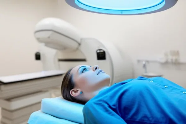
Okay, now let's see what happens inside the test. After you're taken into the test room, you'll be asked to lie down on a bed under the ``Gamma camera.'' This camera is a bit big, but it won't touch your body, so there's nothing to be afraid of.

The camera head will move slowly over your body part being examined, or it will stay in one place, taking a series of pictures. It is very important that you remain still for a long time during this time. It is like holding your breath while taking a photo. If you move, the pictures may not be clear, and you may need to repeat the test. So try to stay still as instructed by the staff. You may sometimes be asked to hold your breath for a while.

Special sensitive crystals inside the camera head pick up the subtle radiation coming from the radioactive tracer in your body. They then create images on a computer screen. Doctors analyze these images to make a diagnosis about your condition.

How long does this test take?

The time it takes for this varies depending on the type of test you are having. Typically, it takes about 30 to 60 minutes just to do the scan with the camera, not including the time it takes for the radioactive tracer to be administered and for it to be absorbed properly.

However, there are some tests, such as bone scans, where it can take two to three hours for the radioactive tracer to be fully absorbed into the body. In such cases, you will be given the tracer, given a short break (perhaps told to drink water during this time), and then asked to come back for the scan at the scheduled time.

There are times when some `(Nuclear Medicine)` tests may need to be done over several days. For example, there may be times when you are given a `(tracer)` today, and then you may have one scan today, and another scan tomorrow. You will be informed of all of this in advance.

What are the risks and benefits of this?

Many people get a little nervous when they hear the word 'radioactive', and that's normal. But as I said before, the tracers used in these tests are not drugs, and they usually don't cause side effects. Also, the level of radiation you receive during a test like this is very low, so you don't need to worry about things like cancer risk.

However, very rarely, that is, in a very small percentage, some people may have an allergic reaction to the `(Radioactive tracer)`. That means something like a rash or itching on the body. Therefore, if you are allergic to anything (such as medicines, foods, dust), you should definitely tell your doctor and the people who are doing the test about it in advance. Then they will be attentive to it.

Now let's look at the advantages. These `(Nuclear Medicine)` scans can provide us with very important and unique information that we cannot get from other tests. Sometimes, something that needs to be done for major surgery (`exploratory surgery`) can be found with a scan like this, without causing much harm to the body (`least-invasive`). That is, this is a great help in improving the quality of diagnosis and treatment. In many cases, it can also detect diseases in their early stages, even at subtle stages that are not caught by other tests. This provides a great opportunity to start treatment early.

After the test?

Usually, after the test is over, you can go back to your normal activities, eat and drink your normal food. No special rest is needed. Sometimes the doctor will tell you to drink a little more water, to help the ``(tracer)`` leave your body more quickly. Other than that, there is nothing special. The doctor will talk to you about it when you get your test report.

Finally, the most important things to remember (Take-Home Message)

Okay, we've talked a lot about ``Nuclear Medicine Imaging'' now. I think you've got a good understanding of it. Here are the main things you need to remember:

  • Nuclear Medicine tests are a highly advanced, safe, and effective method used to diagnose many diseases and treat some.
  • These do not cause much harm or pain to the body (`least-invasive`). The amount of radiation is also very low.
  • If your doctor tells you about this type of test, talk to him or her about it. Ask him or her about any questions or concerns you have. Don't hold anything back.
  • Follow the instructions exactly, especially regarding preparation before the test.

"Remember, with the advancement of medical science, tests like this are very helpful in protecting our health and detecting diseases quickly. Therefore, it is important to know the correct information and follow medical advice without creating unnecessary fear."

If you want to know anything else, be sure to ask your doctor. Stay healthy!

👩🏽‍⚕️ Additional questions (FAQs)

💬 What is a Nuclear Medicine Imaging scan?

Instead of looking at the body with a light from outside like an X-ray, a special sugary liquid (tracer) that produces radiation is injected into our body. Then, a special camera (gamma camera) is used to record video from outside to see how the injected radiation travels inside our body and which organs absorb it.

💬 Is this a dangerous scan with deadly radiation?

Although the name may sound scary, it only uses a very small dose of radiation (tracer). The radiation that enters the body from this is no different than taking a few regular X-rays, and it is completely eliminated from the body in the urine within a few hours.

💬 What diseases is this scan most often used to detect?

This nuclear liquid method is used daily around the world to see the functioning of Thallium Scan (stress test), which is essential to check if there is not enough blood flowing to the heart muscle, PET scan, which looks for the smallest cancer cells, and Thyroid scans, which are used to clearly image thyroid tumors.


` Nuclear medicine, radiological tests, diagnostics, tracers, gamma cameras, health, medical tests

⚠️ Important: The medical articles and information on Nirogi Lanka are for general awareness only, and are by no means a substitute for professional medical advice, diagnosis, or treatment. For any medical problem you have, consult a qualified physician immediately.

💬 Comments (0)

No comments yet. Be the first to share your thoughts here.

Add Your Comment

Please calculate: 2 + 1 =
A special camera that looks inside the body: Everything about Nuclear Medicine Imaging!
How the Body WorksJuly 5, 2026

A special camera that looks inside the body: Everything about Nuclear Medicine Imaging!

Have you ever heard of a test called 'Nuclear Medicine Imaging'? You probably heard about it when your doctor told you to do such a test. It really sounds a little scary when you hear this name, doesn't it? 'Nuclear' when you say it like that. But don't worry, today we will talk about it in detail, in Sinhala that you can understand, and very simply. Just like talking to a friend.

What is this so-called Nuclear Medicine Imaging?

Simply put, this is a special method used to look at the organs inside our bodies. But unlike other scans, this one has a slight difference. Here's what happens: First, you are given a very small amount of a 'radioactive tracer'. This is most often given through an injection into a vein. Sometimes it is given as a drink, depending on the test being performed. This is not a medicine or a liquid that dyes the body, is it? When this is given, it travels through our blood, goes to the organ or tissue that we want to examine, and is deposited there.

Then, you are placed under a special camera called a 'gamma camera', which detects the subtle radiation emitted by the substance that was inserted into your body. This camera is very sensitive. That radiation information is sent to a computer and turned into beautiful images. It's like a 'map' of an organ inside our body. It's by looking at those images that doctors can find out if there is any problem in your body or if an organ is working properly.

Now you might be thinking, 'Oh, radioactive, it's scary, it must be bad for your body.' But the truth is, the amount of radiation in the material used for this test is very low . That is, it is the same amount of radiation that we are usually exposed to in our daily environment, and sometimes even less. Also, these materials usually do not cause any side effects. These materials are eliminated from the body after a while. So there is no reason to be unnecessarily afraid.

Why do we need this type of test (Nuclear Imaging Test)?

Well, this test is mainly used to diagnose various diseases. Sometimes it is also used to treat and see how effective the current treatment is. Just imagine how much this test can help our doctors find. Here are some of the diseases and conditions that this test can detect:

  • Some blood diseases (e.g., some types of anemia, blood clotting problems).
  • Thyroid diseases, especially conditions such as hypothyroidism, where the thyroid gland does not function properly.
  • Heart disease (e.g., reduced blood flow to the heart muscle, damage to the heart after a heart attack).
  • Gallbladder diseases (e.g., cholecystitis, bile duct obstruction).
  • Lung problems (e.g., blood clots in the lungs, decreased lung function).
  • Bone-related problems, such as bone infections, hard-to-find fractures, and whether cancer cells have spread to the bones.
  • Kidney diseases, for example, kidney infections, scarring, and urinary tract obstruction.
  • Identify different types of cancer and see how far the cancer has spread (staging).

In addition to this, as I mentioned earlier, this ``Nuclear Medicine Imaging'' technology is also used for treatment methods. An example is the ``Radioimmunotherapy'' treatment method. In this, radiation and immunotherapy are combined, and radiation is sent to a targeted area, just like cancer cells, for treatment. This means that damage to healthy cells can be minimized.

What is the difference between this and other radiologic tests?

This is a very important question for you to understand. Other `(Radiologic tests)` or as we know it ``X-ray``, ``(CT scan)`` mainly look at how the organs of our body look , that is, their shape, size, density, structure, etc. Doctors call this looking at the ``anatomy`` of an organ.

But this ``Nuclear Medicine Imaging'' test mainly looks at how the organs are functioning . That is, whether the organs are working properly, whether their chemical processes are occurring properly, whether blood circulation is good, and whether there are any changes at the cellular level.

To be precise, while other scans take a 'photo' of an organ, Nuclear Medicine Imaging is like 'observing live how that organ works'.

The biggest advantage of being able to see the function of an organ in this way is that some diseases can be identified by changes in their function before there are major changes in their structure. Then doctors can catch the disease at an early stage and start treatment quickly. This increases the chance of treatment being successful.

How should we prepare before this test?

This is also something you need to know for sure. But since there are so many types of ``Nuclear Medicine'' tests, the way you need to prepare for each one is not the same. It will vary from test to test that you are scheduled to do.

For example, for some tests, you may be asked not to eat or drink anything except water for four to six hours before the test until the test is over (fasting). For other tests, you may be asked to stop taking certain medications a few days before the test. But other tests may not require any special preparation.

Therefore, the most important thing is that when you are prescribed a test like this, you should ask your doctor or the relevant department of the hospital where the test is being performed exactly how to prepare for it. They will give you precise instructions on what to do and what not to do. Following those instructions exactly is very important to get accurate test results.

What should we know on the day of the test?

There are a few little things you need to keep in mind on the day of the test. These aren't big things, but they're good to know.

  • First of all, avoid bringing your valuables (like jewelry, pearls), large amounts of money, and credit cards to the hospital. Because if they are lost, that's another problem. Even though there are usually locker facilities, it's best not to bring them.
  • You may be asked to wear a special hospital gown. This will make the test easier.
  • This test is performed by specially trained, registered technologists. The images and data obtained from the test are then analyzed and diagnosed by Nuclear Radiologists and physicians. So, you can be sure that you are in safe hands.

What happens during the test?

Okay, now let's see what happens inside the test. After you're taken into the test room, you'll be asked to lie down on a bed under the ``Gamma camera.'' This camera is a bit big, but it won't touch your body, so there's nothing to be afraid of.

The camera head will move slowly over your body part being examined, or it will stay in one place, taking a series of pictures. It is very important that you remain still for a long time during this time. It is like holding your breath while taking a photo. If you move, the pictures may not be clear, and you may need to repeat the test. So try to stay still as instructed by the staff. You may sometimes be asked to hold your breath for a while.

Special sensitive crystals inside the camera head pick up the subtle radiation coming from the radioactive tracer in your body. They then create images on a computer screen. Doctors analyze these images to make a diagnosis about your condition.

How long does this test take?

The time it takes for this varies depending on the type of test you are having. Typically, it takes about 30 to 60 minutes just to do the scan with the camera, not including the time it takes for the radioactive tracer to be administered and for it to be absorbed properly.

However, there are some tests, such as bone scans, where it can take two to three hours for the radioactive tracer to be fully absorbed into the body. In such cases, you will be given the tracer, given a short break (perhaps told to drink water during this time), and then asked to come back for the scan at the scheduled time.

There are times when some `(Nuclear Medicine)` tests may need to be done over several days. For example, there may be times when you are given a `(tracer)` today, and then you may have one scan today, and another scan tomorrow. You will be informed of all of this in advance.

What are the risks and benefits of this?

Many people get a little nervous when they hear the word 'radioactive', and that's normal. But as I said before, the tracers used in these tests are not drugs, and they usually don't cause side effects. Also, the level of radiation you receive during a test like this is very low, so you don't need to worry about things like cancer risk.

However, very rarely, that is, in a very small percentage, some people may have an allergic reaction to the `(Radioactive tracer)`. That means something like a rash or itching on the body. Therefore, if you are allergic to anything (such as medicines, foods, dust), you should definitely tell your doctor and the people who are doing the test about it in advance. Then they will be attentive to it.

Now let's look at the advantages. These `(Nuclear Medicine)` scans can provide us with very important and unique information that we cannot get from other tests. Sometimes, something that needs to be done for major surgery (`exploratory surgery`) can be found with a scan like this, without causing much harm to the body (`least-invasive`). That is, this is a great help in improving the quality of diagnosis and treatment. In many cases, it can also detect diseases in their early stages, even at subtle stages that are not caught by other tests. This provides a great opportunity to start treatment early.

After the test?

Usually, after the test is over, you can go back to your normal activities, eat and drink your normal food. No special rest is needed. Sometimes the doctor will tell you to drink a little more water, to help the ``(tracer)`` leave your body more quickly. Other than that, there is nothing special. The doctor will talk to you about it when you get your test report.

Finally, the most important things to remember (Take-Home Message)

Okay, we've talked a lot about ``Nuclear Medicine Imaging'' now. I think you've got a good understanding of it. Here are the main things you need to remember:

  • Nuclear Medicine tests are a highly advanced, safe, and effective method used to diagnose many diseases and treat some.
  • These do not cause much harm or pain to the body (`least-invasive`). The amount of radiation is also very low.
  • If your doctor tells you about this type of test, talk to him or her about it. Ask him or her about any questions or concerns you have. Don't hold anything back.
  • Follow the instructions exactly, especially regarding preparation before the test.

"Remember, with the advancement of medical science, tests like this are very helpful in protecting our health and detecting diseases quickly. Therefore, it is important to know the correct information and follow medical advice without creating unnecessary fear."

If you want to know anything else, be sure to ask your doctor. Stay healthy!

👩🏽‍⚕️ Additional questions (FAQs)

💬 What is a Nuclear Medicine Imaging scan?

Instead of looking at the body with a light from outside like an X-ray, a special sugary liquid (tracer) that produces radiation is injected into our body. Then, a special camera (gamma camera) is used to record video from outside to see how the injected radiation travels inside our body and which organs absorb it.

💬 Is this a dangerous scan with deadly radiation?

Although the name may sound scary, it only uses a very small dose of radiation (tracer). The radiation that enters the body from this is no different than taking a few regular X-rays, and it is completely eliminated from the body in the urine within a few hours.

💬 What diseases is this scan most often used to detect?

This nuclear liquid method is used daily around the world to see the functioning of Thallium Scan (stress test), which is essential to check if there is not enough blood flowing to the heart muscle, PET scan, which looks for the smallest cancer cells, and Thyroid scans, which are used to clearly image thyroid tumors.


` Nuclear medicine, radiological tests, diagnostics, tracers, gamma cameras, health, medical tests

⚠️ Important: The medical articles and information on Nirogi Lanka are for general awareness only, and are by no means a substitute for professional medical advice, diagnosis, or treatment. For any medical problem you have, consult a qualified physician immediately.

💬 Comments (0)

No comments yet. Be the first to share your thoughts here.

Add Your Comment

Please calculate: 2 + 1 =