

You wake up after a solid night's rest, look in the bathroom mirror, and notice them right away: shadowed, purplish, or deep brownish rings beneath your lower eyelids. Almost everyone has had a coworker, friend, or relative ask, "Are you feeling tired today?" when, in reality, you feel perfectly alert. This experience is remarkably common across all ages and ethnicities, yet it remains one of the most frustrating aesthetic concerns encountered in dermatological practice.
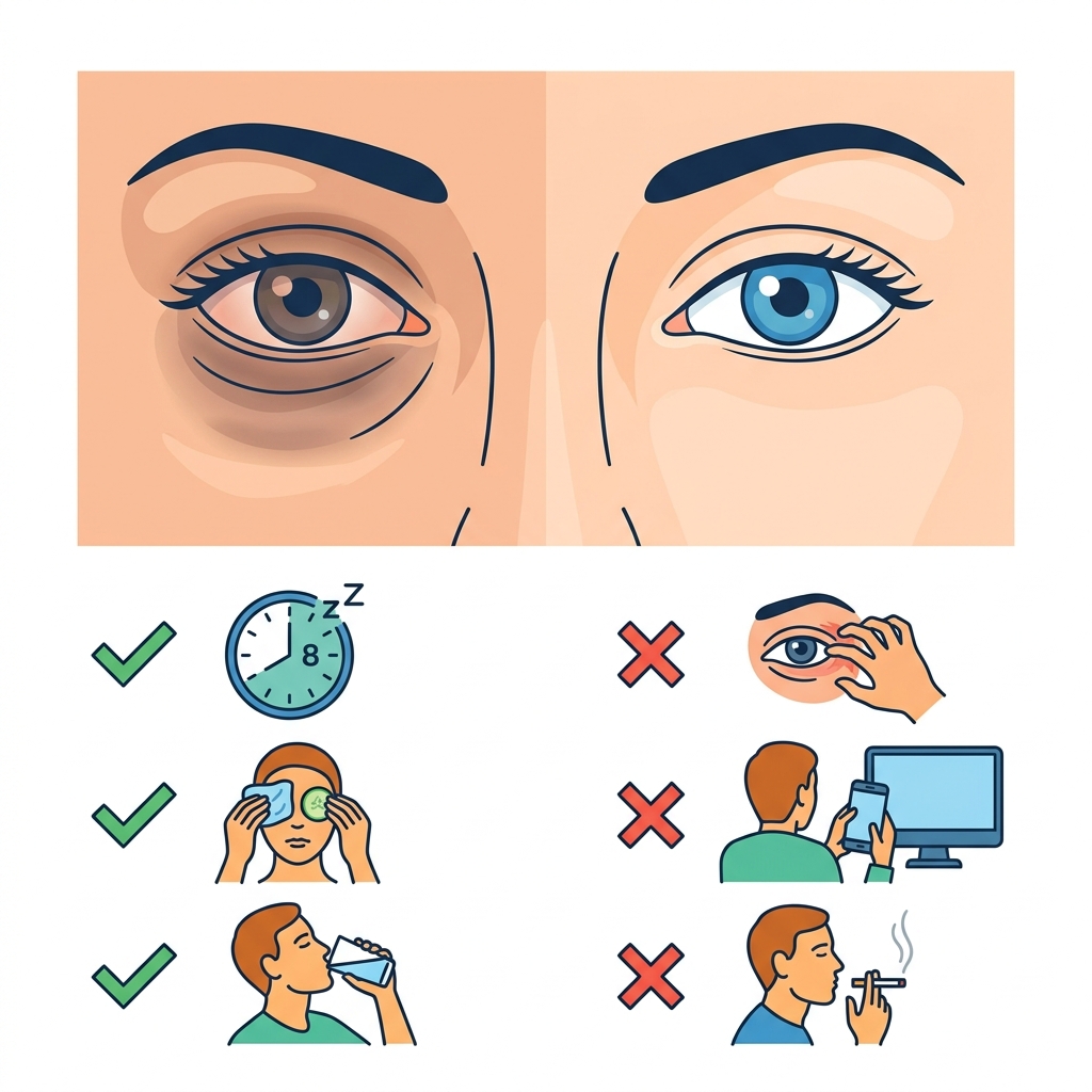
Known clinically as periorbital hyperpigmentation or infraorbital dark circles, this condition rarely signals an underlying systemic disease. However, because the tissue beneath our eyes is the thinnest on the human body—measuring roughly 0.5 mm in thickness compared to 2.0 mm across the rest of the face—every physiological shift in blood flow, pigment deposition, and subcutaneous fat becomes amplified. Understanding the precise anatomical reason for your dark circles is the first critical step toward finding a solution that actually works.
⚡ Key Clinical Takeaways.
- Multifactorial Etiology: Dark circles are rarely caused by fatigue alone. They stem from a combination of hereditary dermal hyperpigmentation, structural shadowing (tear troughs), and thin skin revealing underlying venous networks.
- Allergic Shiners: Chronic seasonal allergies and atopic dermatitis dilate periorbital venules and provoke repetitive eye-rubbing, leading to secondary post-inflammatory hyperpigmentation.
- Tailored Interventions: Treatment must match the anatomical subtype: topical depigmenting agents for excess melanin, volume restoration (dermal fillers) for structural hollowing, and vascular lasers or cold therapy for prominent venous pools.
- When to Seek Urgent Care: Bilateral infraorbital darkness is typical, but sudden unilateral (one-sided) swelling, progressive discoloration, bruising following head trauma, or proptosis (eye bulging) warrants prompt medical evaluation.
BMI Calculator
Know your body mass index (BMI) and weight classification
What Causes Dark Circles Under Your Eyes?
Dermatologists and oculoplastic surgeons classify periorbital dark circles into distinct clinical categories based on their primary anatomical mechanism. Many patients present with a "mixed" type, involving two or more overlapping factors.
1. Structural Anatomy and Age-Related Volume Loss.
As we age, the body gradually slows the synthesis of structural type I and III collagen and elastin. Concurrently, the infraorbital fat pads atrophy, and the underlying orbital bone undergoes slow remodeling and resorption. This anatomical shifting deepens the tear trough (the groove that runs from the inner corner of the eye down across the upper cheek). When overhead ambient lighting strikes these hollowed depressions, it creates an optical shadow that gives the illusion of dark, sunken skin, even when the skin itself contains no excess pigment.
2. Vascular Pooling and Dermal Thinning.
The skin overlying the orbicularis oculi muscle is exceedingly delicate. If microcirculation in the infraorbital venous plexus slows down—whether due to lack of restorative sleep, nasal mucosal congestion, or genetics—deoxygenated blood pools beneath the lower lids. Through translucent, paper-thin skin, this pooled venous blood reflects light with a bluish or purplish tint, often accompanied by mild fluid retention (periorbital edema).
3. Melanin Hyperdeposition (Hyperpigmentation).
Excessive production and deposition of melanin within the basal and suprabasal layers of the epidermis or dermis leads to brown or slate-gray discoloration. This presentation is especially prevalent among people of Mediterranean, Middle Eastern, South Asian, and African ancestry. It can be triggered or exacerbated by chronic ultraviolet (UV) solar radiation, hormonal fluctuations, and persistent mechanical friction.
4. Chronic Allergies and "Allergic Shiners".
Patients suffering from allergic rhinitis, asthma, or atopic dermatitis often develop dark, bruised-looking infraorbital crescents known in clinical pediatrics and immunology as allergic shiners. Histamine release during an allergic flare triggers widespread vasodilation and capillary engorgement in the nasal mucosa and surrounding periorbital veins. also, the persistent itchiness prompts frequent, aggressive eye rubbing, which damages microcapillaries and deposits hemosiderin while inducing localized post-inflammatory hyperpigmentation (PIH).
5. Lifestyle and Systemic Influences.
While lifestyle habits are rarely the sole driver of persistent dark circles, they noticeably modulate their severity:
- Suboptimal Hydration: Dehydration reduces overall tissue turgor, causing the skin beneath the eyes to pull inward against the orbital rim, accentuating hollows.
- Chronic Sleep Deprivation: Prolonged wakefulness elevates endogenous cortisol levels, which diverts blood flow away from the cutaneous microcirculation, leaving the facial skin pale and maximizing the contrast of underlying blood vessels.
- Tobacco and Excessive Alcohol Consumption: Nicotine accelerates collagen degradation and degrades microvascular endothelial function, while excessive alcohol promotes peripheral vasodilation and systemic fluid imbalance.
🚨 When to Seek Emergency Medical Attention.
While standard bilateral dark circles are benign cosmetic concerns, distinct periorbital symptoms can herald acute ocular, neurological, or trauma-related emergencies. Call 911 (US/Canada), 999 (UK), or proceed directly to your nearest Emergency Department (ER / A&E) if you experience:
- Raccoon Eyes (Periorbital Ecchymosis): Bilateral purplish bruising around both eyes appearing hours or days after a head injury or fall, which is a classic sign of a basilar skull fracture.
- Acute Unilateral Swelling with Severe Pain and Fever: Redness, heat, deep pain with eye movement, or proptosis (bulging eye), indicating potential orbital cellulitis or cavernous sinus thrombosis.
- Sudden Visual Disturbances: Blurred vision, double vision (diplopia), loss of peripheral vision, or an inability to move your eyeball properly.
- Rapidly Progressive Unilateral Discoloration: Unexplained, expanding dark pigmentation confined to a single eyelid that bleeds, crusts, or changes shape, warranting urgent evaluation to exclude malignant melanoma.
Home Remedies and Daily Self-Care: What Actually Works?
Conservative, non-invasive habits can reduce periorbital puffiness, constrict congested capillaries, and improve superficial skin barrier health:
- Targeted Cold Compresses: Applying a chilled gel eye mask, clean cold spoons, or ice wrapped in a soft cloth for 5 to 10 minutes constricts dilated superficial venules via localized vasoconstriction, visibly decreasing immediate bluish undertones and morning puffiness.
- Caffeinated Cold Tea Bags: Chilled steeped green or black tea bags contain high concentrations of caffeine and anti-inflammatory polyphenolic catechins (such as epigallocatechin gallate, or EGCG). Topical caffeine penetrates the thin stratum corneum to stimulate local microcirculation and temporarily constrict lymphatic and vascular vessels.
- Elevation During Sleep: Sleeping with the head slightly elevated by using an extra ergonomic pillow prevents fluid from pooling in the loose infraorbital connective tissue overnight, minimizing puffy morning bags that cast secondary shadows.
- Broad-Spectrum Sun Protection: Diligently applying an ophthalmologist-tested mineral sunscreen (zinc oxide or titanium dioxide, minimum SPF 30) daily around the periorbital margin prevents UV-induced melanogenesis and protects dermal collagen.
- Targeted Topical Actives: Formulations containing vitamin C (L-ascorbic acid) brighten hyperpigmented skin and neutralize free radicals; retinol or retinaldehyde stimulates steady neocollagenesis over months; and hyaluronic acid transiently plumps surface fine lines.
Medical and Dermatological Interventions.
When dark circles do not respond to topicals and lifestyle modifications, a board-certified dermatologist or plastic surgeon can assess your specific anatomical subtype and recommend clinical procedures:
| Clinical Procedure. | Mechanism of Action. | Best Indication & Recovery. |
|---|---|---|
| Hyaluronic Acid Fillers . | Gel injected deep along the periosteum restores lost infraorbital volume, smoothing the transition from lower lid to cheek and eliminating the shadow gap. |
Ideal for: Structural tear trough hollows. Note: Requires an experienced injector to avoid the Tyndall effect (a bluish tint caused by superficial product placement). Minimal downtime. |
| Prescription Depigmenting Agents . | Formulations using hydroquinone (2% to 4%), azelaic acid, kojic acid, or tranexamic acid selectively inhibit tyrosinase, the rate-limiting enzyme in melanin synthesis. |
Ideal for: True pigmentary dark circles. Note: Must be applied carefully under medical guidance to avoid paradoxical ochronosis or periorbital dermatitis. |
| Chemical Peels . | Superficial application of glycolic, lactic, trichloroacetic (TCA), or mandelic acid exfoliates superficial keratinocytes and lifts trapped epidermal pigment. |
Ideal for: Superficial epidermal hyperpigmentation. Note: 2 to 5 days of mild flaking and erythema. |
| Vascular and Pigment Lasers . | Pulsed dye lasers (PDL) target visible red/purple venules, while Q-switched or picosecond lasers shatter deeper dermal melanin deposits. Fractional resurfacing lasers thicken collagen. |
Ideal for: Prominent superficial microvessels and severe dermal melasma. Note: Requires corneal eye shields; slight bruising for 3 to 7 days. |
| Platelet-Rich Plasma (PRP) . | Autologous plasma concentrated with platelets is micro-injected into the dermis, releasing growth factors (VEGF, PDGF, TGF-beta) to stimulate localized tissue remodeling. |
Ideal for: Thin, crepey lower eyelid skin. Note: Usually requires 3 sessions spaced 4 to 6 weeks apart; transient swelling for 24–48 hours. |
| Lower Blepharoplasty . | A surgical procedure performed via transconjunctival or subciliary incision to reposition herniated orbital fat into the tear trough groove and excise redundant skin. |
Ideal for: Prominent lower eye bags causing harsh, fixed shadows. Note: Permanent structural correction; 1 to 2 weeks of post-op ecchymosis. |
Do's and Don'ts for Periorbital Care.
To avoid irritating the infraorbital region and accelerating skin aging, follow these daily evidence-based guidelines:
| Do's (Protective Habits). | Don'ts (Aggravating Habits). |
|---|---|
| Treat Allergies Proactively: Manage allergic rhinoconjunctivitis using daily non-sedating oral antihistamines (cetirizine, fexofenadine) or prescribed intranasal corticosteroid sprays to stop nasal congestion and eye itching. | Do Not Rub or Scrub Your Eyes: Aggressive rubbing damages tiny capillaries, leading to micro-bleeding, hemosiderin staining, and long-term lichenification with darkening of the skin. |
| Wear UV-Blocking Sunglasses: Choose large, wraparound lenses with broad UV400 protection. This blocks melanocyte activation and stops you from squinting, which strains periorbital muscles. | Do Not Apply Harsh Body Bleaches: Never use unregulated high-strength hydroquinone, ammoniated mercury compounds, or heavy body exfoliants near the eyes. This can cause chemical burns, severe eczema, or permanent ocular injury. |
| Remove Makeup Gently: Use a fragrance-free micellar water or oil-based cleanser, soaking a soft cotton pad and pressing it gently against the eye to dissolve makeup without mechanical tugging. | Avoid High-Sodium Late-Night Dinners: Heavy sodium intake prior to sleep forces renal osmotic fluid retention, precipitating severe morning periorbital edema and venous stasis. |
| Perform the "Pinch Test": Gently lift the infraorbital skin. If the pigment lifts and stays dark, it is true melanin; if the darkness disappears when the skin is elevated away from the underlying muscle, it is vascular or shadow-based. | Don't Expect Eye Creams to Fix Hollows: Topicals cannot restore lost bone or deep fat pads. Relying solely on creams for severe structural tear troughs leads to unnecessary expense and disappointment. |





No comments yet. Be the first to share your question or experience below.