

Do you have a hard time going to the toilet? Or do you sometimes have trouble holding your bowels in? Or do you feel strange, unfamiliar discomfort when you go to the toilet? If you have any of these problems, your doctor may want you to have a special test called a defecography to find out the cause. Simply put, this is done to look at how the muscles and other organs inside your body work when you have a bowel movement.
What is Defecography?
Okay, let's take a closer look at what defecography is. This is actually a radiology test. It looks at how the muscles and organs inside your body are working when you defecate, or as doctors call it, "defecate." This is done using a special "Fluoroscopic X-ray" test or an "MRI - Magnetic Resonance Imaging" test. Through these technologies, doctors can clearly see how different parts of your body are moving and how they are functioning when you defecate.
Now you may be wondering if you really have to poop for this test. No, you don't. The medical staff will put a special paste that is exactly like poop in your rectum. It's called a "barium" paste. This "barium" is a "contrast agent," a substance that makes the inside of your body show up clearly on an "X-ray" or "MRI." You will then pass out this paste while sitting in a special toilet inside the scanner. The important thing is that the radiologist is not looking at you on the toilet, but at the images of your insides on the computer screen. So there's nothing to be embarrassed about.
When is this test needed?
The main purpose of this test is to let your doctor know more about what's happening inside when you poop. This will help them find the exact cause of your problem and provide the right treatment. This test helps doctors find out about things like:
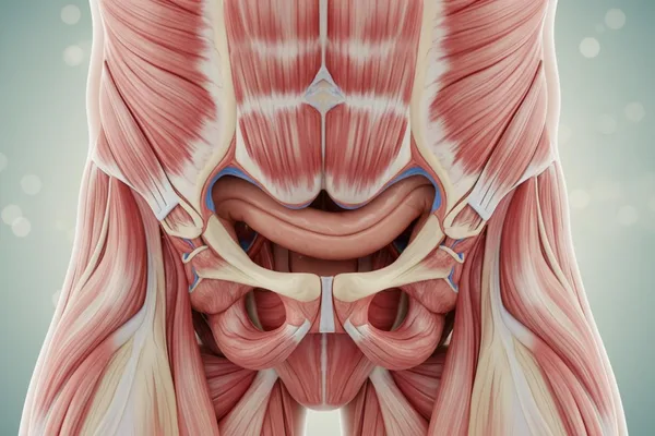
- The way your pelvic floor muscles contract and relax.
- How your anus works when you have a bowel movement.
- The way your colon, rectum, vagina, and bladder move when you defecate.
- Has any organ inside your body shifted or changed position?
- Perhaps if you're planning to have surgery, what kinds of things should you be especially careful about?
What diseases can be detected by this test?
If you have unexplained symptoms related to your bowel movements for a long time, your doctor may recommend a defecography test to find the cause. For example:
- Anal pain.
- Constipation (meaning difficulty in defecating).
- Inability to control the passage of stool (Fecal incontinence).
- Incomplete evacuation is the feeling that the stool has not been completely expelled.
This test is also used to confirm a medical condition if the doctor suspects it. Such conditions include:
- Pelvic floor dysfunction: Simply put, the muscles and nerves in your pelvic floor do not coordinate and function properly when you defecate.
- Organ prolapse: This is when an organ in your body weakens and falls out of place, or one organ pushes into another. Some examples are:
- Rectal prolapse.
- Rectocele is the protrusion of the rectum into the vagina.
- Enterocele is a protrusion of the small intestine into the anus.
Who is doing this test?
All such X-ray and MRI examinations are performed by specially trained, certified radiologists. Defecography, in particular, is performed by a specialist with a deep understanding of the rectum and its associated movements (Anorectal Motility).
How is this test done?
Defecography can be done using either a fluoroscopic X-ray or an MRI. Both methods can look at how your body's internal mechanisms work when you defecate. The X-ray method is the most commonly used, but for some people, the small amount of radiation emitted by an X-ray can be harmful, so an MRI is more suitable.
Think of it like a video X-ray. Instead of taking pictures at one point, a continuous beam of X-rays is sent through the body. You can then watch a series of moving images on a screen in real-time. That means you don't have to wait for the photos to be developed, you can watch your organs work like a video.
An MRI (Magnetic Resonance Imaging) test works by using radio waves and a magnetic field. An electric current is passed through metal coils throughout the machine to create a magnetic field. This causes the hydrogen atoms in your body to move. When they return to their original positions, the machine picks up the signal they emit through radio waves and displays it as an image on a computer.
What happens before the test?
Your doctor will take a detailed note of your complete medical history, including any surgeries you've had, medical conditions you have, medications you take, and any implants you have. Then, they will ask you in detail about what exactly happens when you defecate.
Next, we will give you a full explanation of how this test is performed. We will also explain how you can support us to get the clearest results from the test. Because the success of this test largely depends on your support.
Sometimes, you may be given an enema to use at home to help clear your bowels before the test. You may also be asked to change your diet, medications, and fluid intake before the test. These instructions may vary from person to person, so it is important to follow your doctor's instructions exactly.
How do you prepare yourself on test day?
Whether you are having a Fluoroscopic X-ray Defecography or an MRI Defecography, both of these tests require the use of a contrast agent that makes the internal organs that the doctor wants to see clearly. This means that this contrast agent must be injected into your body before the test.
To see the last part of your digestive system – the colon, rectum, and anus – clearly, the medical team will put a contrast agent (Barium Sulfate) in a paste and insert it into your rectum. The Barium Enema is given right after the exam. However, sometimes the doctor may want to look at other organs that the Enema cannot reach.
For example, if you need to see your small intestine, you may be given a contrast solution to drink. This may need to be taken a few hours before the test, because it takes time for the solution to pass through your digestive system. At another time, if you need to see the wall between your vagina and rectum, a barium paste may be applied to your vagina.
What is the experience like during a defecography test?
After you put on a hospital gown, you will be taken to the examination room. There, you will lie down on a table. Then, the medical team will insert a barium paste into your rectum using a caulking gun. This enema may make you feel a little full or a little bloated. When your rectum is full, you will naturally feel an urge to defecate. You are then ready for the test.
If you are in an X-ray machine or an open MRI machine, the table you are on is tilted vertically, and there is a special toilet under you. You usually sit on the toilet. Both of these machines are designed to be claustrophobic, with plenty of space and ventilation. But if you are in a traditional MRI machine, which is a bit like a tube, you have to lie flat on the table, and then you have to defecate into an adult diaper. You may also be covered with a blanket with metal coils.
Some people may feel claustrophobic when they are inside the traditional MRI machine. If this happens, you may be given a sedative to help you relax. The machine is well-lit and air-conditioned. You can also talk to the technician through an intercom. Both open and traditional MRI machines make a loud "tock tock, click click" sound when the images are taken. You will be given earplugs or headphones before the procedure.
The radiologist who operates the test equipment will give you instructions from outside the machine. As they take the images, they will tell you to tense, hold, and relax your muscles. When you push, you need to apply enough force so that the technician can clearly see how your muscles are working. You also need to remain very still while the images are taken continuously – usually for less than a minute.
The entire test takes about 30 to 60 minutes. It may feel a little tiring and maybe even embarrassing, but it is not a painful test.
What happens after the test?
After the test is over, you will be taken to a private bathroom where you can clean yourself. You will likely see a white, chalky substance called ``Barium'' pass out of your stool for about two days. You will be able to resume your normal activities and eating habits immediately.
Are there any risks or side effects to this test?
The small amount of radiation emitted by an X-ray is considered safe, as long as you are not exposed to it frequently. An MRI scan does not pose any risks or side effects. If you are pregnant, it is generally recommended to avoid both tests as much as possible, even though the risk of harm to the fetus is very small.
What are the results? What is the meaning of Eve?
The radiologist will send a detailed report to your primary care physician. He or she will tell you the results. The report will reveal whether there are any common problems with bowel movements. It will also tell you if your primary care physician has ordered any specific tests.
Imagine that your doctor has ordered this type of defecation test, meaning that you may have been suffering from this problem for a long time. So, the information from your defecography test is essential for your doctor to develop the best treatment plan to improve your defecation experience.
Take home message
Yes, this test may seem a little strange, and maybe even uncomfortable. But remember, it will help you find the exact cause of your problem and start a suitable treatment for it. In the end, this test can bring you great relief. Therefore, if your doctor recommends this test, it is important to talk about it thoroughly and get it done if necessary.
` Constipation, Difficography, Difficulty defecating, Barium, X-ray, MRI, Pelvic floor




💬 Comments (0)
No comments yet. Be the first to share your thoughts here.
Add Your Comment