
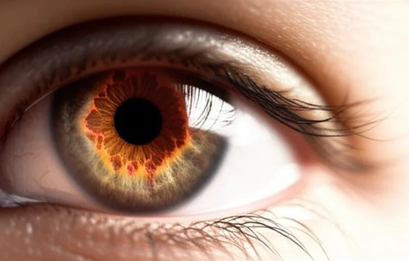
Have you ever wondered how an eye doctor looks at the tiny, yet very important things inside our eyes? Sometimes they put medicine in the eye and make the black eye look bigger, right? Well, today we are going to talk about a method that takes a photo of the back of the eye, the 'fundus'. This is what we call ``Fundus Photography'' .
What is Fundus Photography?
Simply put, this ``(Fundus Photography)`` is when your eye specialist takes a picture of the back wall of your eye, which is the part we call the 'fundus'. By looking at these pictures, the doctor can examine several parts of the eye that are very important for your vision. This is a very simple, non-invasive test.
What are the important parts inside the eye?
There are several very important structures inside and behind our eyes. It's like in our house, everything has its own place. Think about it, these are the main places:
- Retina: This is the layer that contains the light-detecting cells, called photoreceptors. These cells send signals to the brain about the light they detect. The brain uses these signals to create the big picture you "see."
- Macula: This is the most sensitive part of your retina. It contains photoreceptors that help you see color and fine details. It's like an HD camera! When you read a book, the macula helps you see the text clearly.
- Optic Nerve: This is like the 'data cable' that connects your retina to your brain. Many eye diseases can affect the part of this nerve that connects to the eye.
- Choroid: This layer lies just beneath your retina. It is part of the middle layer of the wall around your eye called the uvea. The blood vessels that supply blood to your retina are located in the choroid.
When is this fundus photography test performed?
This ``Fundus Photography`` test can be done as part of your ``Routine Eye Exam``. Or, if you have any eye symptoms or vision changes, it is also used as part of a special eye exam to find the cause. Special tests like these help find specific signs of eye diseases.
Just think, it is in situations like these that doctors often decide to perform this ``(Fundus Photography)`` test:
- Retinal diseases such as diabetes-related retinopathy or hypertensive retinopathy.
- Injuries or damage to the retina, such as tears or detachments.
- Specific changes in the retina, such as macular degeneration or loss of color vision.
- Uveitis is an inflammatory condition that affects the retina or choroid.
- Optic nerve conditions such as optic atrophy and papilledema.
- Changes or damage to the retina and/or optic nerve due to conditions such as glaucoma.
- Causes of dark spots (Scotomas) in vision or visual field defects.
- Eye cancers and non-cancerous (benign) tumors, such as 'retinoblastoma'.
- Infections that occur inside the eye, such as toxoplasmosis.
How does Fundus Photography work?
This ``Fundus Photography`` test is done using a ``Fundus Camera``. Most eye clinics have at least one of these special devices. Okay, now let's see what these cameras look like.
There are two main types of fundus cameras:
Standard Fundus Cameras
This device looks like a telescope tube. At one end, there is a frame for you to hold your chin and forehead, and at the other end, there is a small lens for the doctor to see through, and there are control buttons. To get the best results from these cameras, the doctor will put pupil-dilating drops in your eyes before the exam. This will allow you to see inside your eye better.
Wide-field Laser-based Cameras
These cameras use special lasers and scanning techniques to quickly take pictures of the fundus, even if your eyes are not dilated (i.e., without the use of medication to enlarge the pupil). However, it is easier to take pictures when your eyes are dilated. These cameras usually have a single circular opening that you can see. That opening is on a large, tall, flat cover. The area around that opening is often covered with a soft rubber covering, so it is comfortable and does not hurt when you hold it against your face.
What to expect before and during the test?
Do you need to prepare before the test?
No, this ``(Fundus Photography)`` test does not require much preparation on your part. There is nothing to worry about. The doctor may put eye drops in your eyes to enlarge the black ring in your eyes before taking the pictures. These drops take a few minutes to work. You just have to be patient for a while.
What happens during the test?
Now, we've talked about two types of fundus cameras. So what you experience will depend on the type of camera the doctor uses. Sometimes, it's possible to take pictures with both types of cameras.
Standard Fundus Cameras
When taking pictures with this equipment, you sit on a chair or stool, facing the frame on your side of the tube. The doctor will clean your forehead and chin with a sanitizing wipe before placing your face.
It's very important to keep your chin and forehead exactly in the frame. That way, it'll be easier for the doctor to take pictures quickly. Once your head is in the right position, you should look into one end of the tube. Inside, you'll see a light shining into your eye.
The doctor sits on the other side of the tube, looks through the eyepiece of the camera, adjusts the camera, points it at specific points, and takes pictures one by one. They tell you to look straight ahead, look right, look left, etc. Depending on the reason for taking the pictures, they take pictures of one or both eyes. Once the pictures are taken, the test is over. Pretty quick, right?
Wide-field Laser-based Cameras
When you look into the aperture of the wide-angle camera, you will see a circular target marker, which you need to look straight at. The background behind the marker may change color. The doctor or technician who is doing the exam will tell you what the colors mean, how to move, and what other things need to be captured in the image.
Once you're in the right position, he'll turn on the camera. You'll see a bright green light, like a light on a sheet of paper inside a copier, pass through your vision. It only lasts a short time, about a second. Don't worry, it won't hurt. Usually, three pictures are taken of each eye. Once the pictures are taken, the test is over.
What happens after the test?
After the fundus images are taken, a doctor or nurse will take you to an exam room. There you will meet an optometrist or ophthalmologist. They will do the rest of the exam, or they will look at your fundus images and explain what they mean.
Are there any risks or side effects to this test?
This `(Fundus Photography)` is a very safe test . There are no side effects. However, if you are given medicine to dilate the pupil of your eye (`(Eye Dilation`), the medicine may cause minor side effects. For example, your vision may be blurred for a while, and your eyes may appear blue outside. But these are usually very minor and will get better after a while. If your doctor has dilated your eyes, your eyes will remain dilated until the medicine wears off. Most of the time, this medicine will wear off within a few hours (your doctor will tell you exactly how long it will take).
If you have photophobia, which means your eyes turn blue when you are outdoors, you may feel a little uncomfortable when the light hits your eyes when taking pictures. This is usually only temporary, and your doctor will help you with that. Most eye exam rooms have dimmable lights, so if you are still uncomfortable with bright light, your doctor will use them.
Can I drive after fundus photography?
Yes, in most cases you can drive without any problems after having ``(Fundus Photography)``. It depends on why you had the pictures taken, the condition of your eyes before the test, and a few other factors.
Many people drive to the doctor's office, and you can drive back after the exam. If the doctor dilated your eyes (used medication to enlarge your blackheads), they may give you disposable shaded covers that you can wear like sunglasses. These are comfortable for your eyes when you go out. If you wear glasses, you can leave these disposable covers on over your glasses until the dilation wears off. However, if you are unsure, it is best to ask your doctor before driving.
What are the results like? What do they tell us?
Fundus cameras in many eye clinics can send images to a computer in the exam room as soon as they are taken. Your doctor can then view the images on a computer screen. They may also show you the images and explain the different things they show. They may say things like, "Look at the optic nerve inside your eye..."
If the fundus of your eye looks normal, your doctor will tell you that. It's reassuring to hear something like , "There's nothing wrong, everything's fine." If the pictures show any significant changes or abnormalities, your doctor will explain them to you. If your doctor has previous pictures of you, he or she may compare them. He or she may say things like, "Here's a little difference from last time."
Based on what we see in the latest photos, he could be doing one or more of these things:
- Recommend further testing (e.g., another special scan)
- Offers treatment options (e.g. medication, laser treatment)
- A follow-up appointment will be scheduled. (For example, let's see you again in 3 months)
- You will be referred to another specialist (e.g. a diabetes specialist).
There's a saying that goes, 'A picture is worth a thousand words.' Just like that, a picture behind your eyes is priceless for the health of your eyes.
Finally, remember this.
So, this `(Fundus Photography)` is a very valuable test to monitor the health of your eyes. Your eye specialist can use these images to see if your eyes are healthy and to identify any problems. What's even better is that this `(Fundus Photography)` test can catch changes inside the eye before you show any symptoms. This is part of a routine eye exam. Sometimes, it can detect diseases that start inside the eye without you even realizing it.
So, you can ask your doctor for more details on how this can benefit you. Don't forget that it's your responsibility to protect your eyes! It's very important to get regular eye exams.
` Fundus Photography, Eye Examination, Retina, Eye Health, Macula, Optic Nerve


💬 Comments (0)
No comments yet. Be the first to share your thoughts here.
Add Your Comment