

Picture a busy municipal water purification plant operating 24 hours a day, testing chemical balances down to the milligram, recycling essential nutrients, maintaining fluid equilibrium, and discharging toxins without missing a beat. Within your own body, this vital operation is carried out quietly by your kidneys and urinary tract. Nestled beneath your ribcage, these two fist-sized organs process roughly 120 to 150 quarts (114 to 142 liters) of blood daily to produce one to two quarts of urine. Because renal injury often progresses silently without early pain, understanding this biological filtration system is one of the most critical steps in safeguarding your long-term health.
Key Clinical Takeaways.
- Multi-System Regulators: Beyond waste removal, kidneys manage blood pressure, produce erythropoietin to stimulate red blood cell production, and activate Vitamin D for mineral metabolism.
- The Functional Unit: Over one million nephrons in each kidney filter metabolic byproducts like urea and creatinine via a delicate, three-stage mechanism: glomerular filtration, tubular reabsorption, and tubular secretion.
- Silent Progression: Chronic Kidney Disease (CKD) can destroy up to 90% of renal function before noticeable symptoms appear, making routine screening (eGFR, urine albumin) essential for at-risk patients.
- Preventive Action: Judicious hydration, limiting nonsteroidal anti-inflammatory drugs (NSAIDs), maintaining blood pressure below 130/80 mmHg, and tight glycemic control preserve renal longevity.
Renal function (eGFR)
Measure kidney function based on creatinine value
What Is the Anatomy of the Kidneys and the Urinary System?
The urinary tract is an integrated drainage system designed to create, transport, store, and evacuate liquid waste. It consists of four major anatomical structures working in continuous sequence:
1. The Kidneys.
Located retroperitoneally on either side of the spine between vertebral levels T12 and L3, the kidneys are bean-shaped organs protected by the lower ribs and surrounded by renal fat pads. Each kidney is divided into an outer cortex and an inner medulla:
- Nephrons: The microscopic filtration units. Each kidney contains about 800,000 to 1.2 million nephrons consisting of a glomerulus (a high-pressure tuft of capillaries) and an intricate tubular system (proximal tubule, loop of Henle, distal tubule, and collecting duct).
- Renal Pelvis: A funnel-shaped basin that collects newly formed urine from the renal calyces and channels it into the ureter.
2. The Ureters.
These two slender, muscular tubes measure about 10 to 12 inches (25 to 30 cm) in length in adults. Rather than relying on simple gravity, the muscular walls of the ureters engage in active, rhythmic peristaltic contractions every 10 to 15 seconds to propel urine downward into the bladder while physiological valves prevent reverse flow (vesicoureteral reflux).
3. The Urinary Bladder.
Located in the pelvis, the bladder is a hollow, distensible organ composed of smooth muscle known as the detrusor muscle. In healthy adults, the bladder comfortably holds 13 to 17 fl oz (400 to 500 mL) of urine. Stretch receptors in the bladder wall trigger the initial sensation to void at about 5 to 7 fl oz (150 to 200 mL).
4. The Urethra.
The conduit that directs stored urine out of the body during micturition (urination):
- Female Anatomy: Measures about 1.5 inches (4 cm) in length and exits just anterior to the vaginal opening. Its short length and proximity to the perineum explain why women experience a substantially higher incidence of ascending urinary tract infections (UTIs).
- Male Anatomy: Measures about 8 inches (20 cm) in length, traversing the prostate gland and the penile shaft, serving a dual role in urinary excretion and reproduction.
How Do Kidneys Form Urine? (The Renal Mechanism).
Urine formation requires three tightly synchronized physiological processes within the nephron:
Step 1: Glomerular Filtration.
Blood enters the kidney via the renal artery under high systemic pressure, flowing into the capillary networks of the glomerulus. The glomerular filtration barrier acts as a physical sieve and electrostatic shield. Small molecules—water, electrolytes (sodium, potassium), glucose, amino acids, and metabolic wastes (urea, creatinine)—pass freely through into Bowman’s capsule, creating glomerular filtrate. Larger structures, such as erythrocytes (red blood cells) and high-molecular-weight proteins like albumin, are kept within the bloodstream.
Step 2: Tubular Reabsorption.
If all filtered fluid were excreted, the human body would rapidly succumb to fatal dehydration and electrolyte depletion within hours. As the filtrate travels through the proximal convoluted tubule and the loop of Henle, active and passive transport mechanisms reabsorb over 99% of the water and virtually 100% of vital filtered nutrients (glucose and amino acids), returning them to the peritubular capillaries.
Step 3: Tubular Secretion.
Simultaneously, capillary blood transfers additional unwanted substances—including hydrogen ions ($H^+$), excess potassium ($K^+$), ammonium, and medicine-based drug metabolites—directly into the tubular fluid. The resulting fluid is concentrated urine, which travels through collecting ducts into the renal pelvis.
Essential Endocrine and Homeostatic Functions Beyond Waste Removal.
Viewing the kidneys solely as waste filters underestimates their complex role in human endocrinology and metabolic homeostasis:
- Blood Pressure Regulation (RAAS Pathway): When renal perfusion drops, specialized juxtaglomerular cells secrete the enzyme renin. Renin converts angiotensinogen to angiotensin I, which is then converted by ACE (angiotensin-converting enzyme) to angiotensin II—a potent vasoconstrictor that also triggers aldosterone release, promoting renal sodium and water retention.
- Erythropoiesis (Red Blood Cell Production): In response to renal tissue hypoxia, interstitial fibroblasts produce erythropoietin (EPO), a glycoprotein hormone that directs the bone marrow to produce mature red blood cells. Chronic renal disease often leads to severe normocytic anemia due to deficient EPO synthesis.
- Calcium and Bone Homeostasis: The kidneys express 1-alpha-hydroxylase, the key enzyme responsible for converting inactive 25-hydroxyvitamin D into biologically active 1,25-dihydroxyvitamin D (calcitriol). Without active calcitriol, the digestive tract cannot efficiently absorb dietary calcium.
- Acid-Base Balance: Kidneys maintain arterial pH within the narrow physiological range of 7.35 to 7.45 by actively reabsorbing filtered bicarbonate ($HCO_3^-$) and excreting excess metabolic protons ($H^+$).
🚨 Red Flags: When to Seek Emergency Medical Attention.
Certain urinary tract and renal signs point to life-threatening emergencies, such as severe urosepsis, acute urinary retention, or sudden renal failure. Call 911 (US/Canada), 999 (UK), or proceed directly to the nearest Emergency Room (A&E) if you develop:
- Frank Hematuria with Clots: Passing gross, bright-red blood or solid clots in your urine.
- Suspected Urosepsis: Burning urination or back pain accompanied by high fever, shaking chills, confusion, extreme lethargy, or rapid heart rate.
- Acute Urinary Retention: Complete inability to void despite a painfully distended, tight lower abdomen.
- Uncontrolled Renal Colic: Agonizing, sudden flank or groin pain accompanied by persistent vomiting and an inability to find a comfortable position (concerning for an obstructing nephrolithiasis).
- Uremic Signs / Anuria: Passing zero urine for over 12 hours, accompanied by shortness of breath, acute confusion, or sudden swelling of the face, hands, and legs.
Common Pathologies Affecting the Kidneys and Urinary Tract.
1. Urinary Tract Infections (UTIs) & Pyelonephritis.
Most commonly caused by uropathogenic Escherichia coli originating from the GI tract, UTIs begin as lower tract infections (cystitis) characterized by dysuria (burning), urgency, and frequency. If untreated, bacteria can ascend via the ureters to infect the renal parenchyma (acute pyelonephritis), presenting with flank pain, high fever, and costovertebral angle tenderness.
2. Nephrolithiasis (Kidney Stones).
Mineral crystallization occurs when urine becomes supersaturated with calcium oxalate, uric acid, or struvite. Stones migrating through the narrow ureter cause intense, spasmodic flank pain that radiates to the groin, often accompanied by microscopic or gross hematuria.
3. Chronic Kidney Disease (CKD).
Defined by structural damage or an estimated glomerular filtration rate (eGFR) below 60 mL/min/1.73 m² persisting for more than three months. The two primary driving etiologies worldwide are diabetic nephropathy and hypertensive nephrosclerosis, which cause microvascular glomerular scarring.
Clinical Diagnostic Framework.
Clinicians assess renal and urinary performance using standardized laboratory and imaging tools:
- Urinalysis (Dipstick & Microscopy): Checks for proteinuria, microhematuria, leukocyte esterase, nitrites (bacterial indicators), and cellular casts.
- Serum Creatinine & Blood Urea Nitrogen (BUN): Creatinine is a breakdown product of muscle metabolism. Normal reference ranges typically span 0.7 to 1.3 mg/dL (62 to 115 µmol/L) for adult males and 0.55 to 1.02 mg/dL (49 to 90 µmol/L) for adult females.
- eGFR (Estimated Glomerular Filtration Rate): Calculated from serum creatinine, age, and sex; values above 90 mL/min/1.73 m² indicate normal function, while values below 15 indicate established kidney failure (Stage 5 CKD).
- Urine Albumin-to-Creatinine Ratio (uACR): Detects early glomerular endothelial leak. A normal value is under 30 mg/g (under 3 mg/mmol); persistent values above 300 mg/g indicate severe albuminuria.
- Renal Ultrasonography: Non-invasive imaging measuring kidney dimensions, identifying obstructive hydronephrosis, cysts, and calculi.
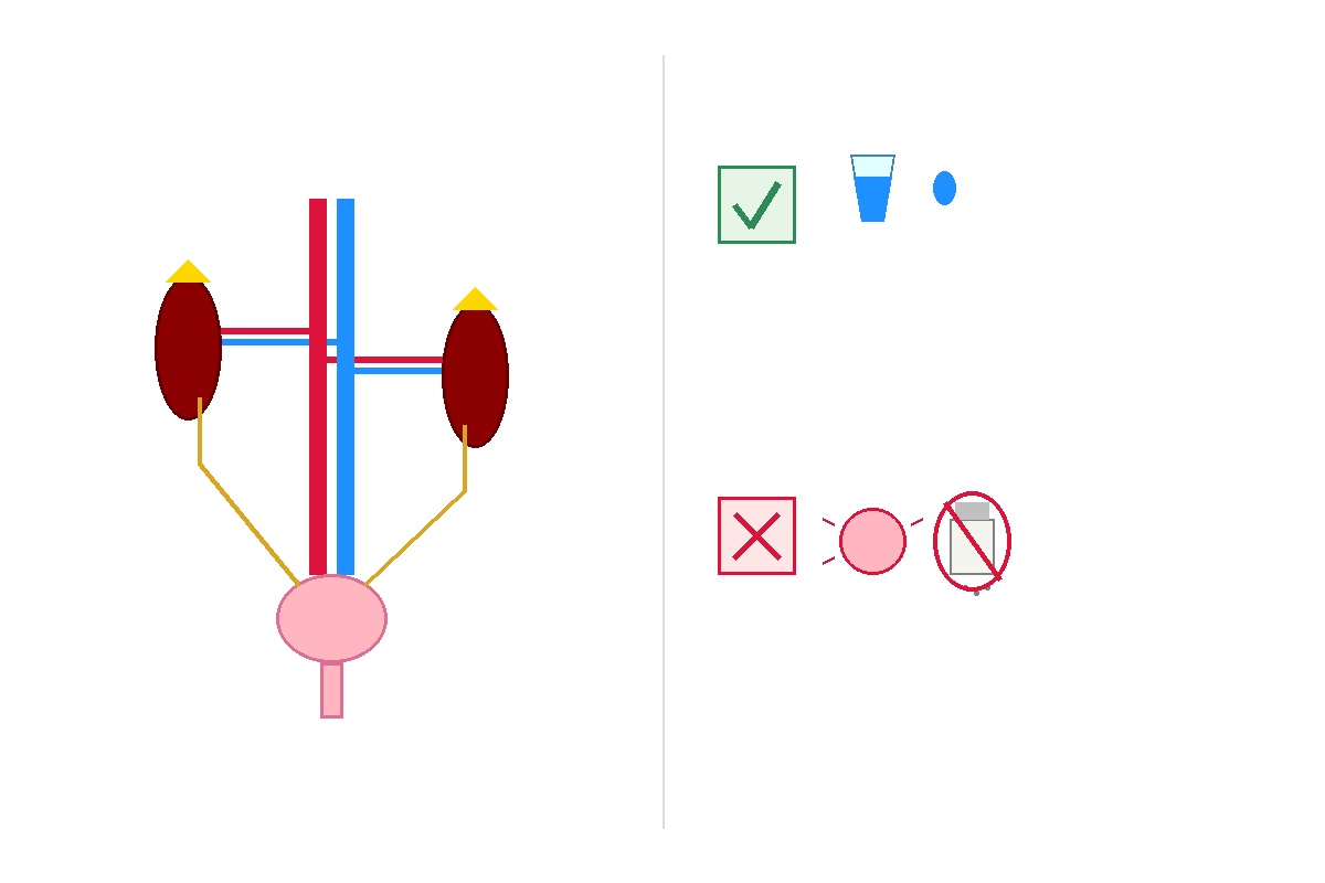
Evidence-Based Guidelines: Do's and Don'ts for Renal Longevity.
| Evidence-Based Practice (DO). | Nephrotoxic Risk / Practice (DON'T). |
|---|---|
| Hydrate Appropriately: Drink enough fluids so your urine remains pale straw-colored. Aim for about 2 to 3 liters (68 to 100 fl oz) of water daily unless medically fluid-restricted. | Don't Overuse NSAIDs: Frequent, unmonitored use of nonsteroidal anti-inflammatory drugs (ibuprofen, naproxen, high-dose aspirin) constricts afferent arterioles, causing acute or chronic interstitial injury. |
| Target Blood Pressure < 130/80 mmHg: Managing systemic pressure shields fragile glomerular capillary beds from hypertensive shear stress. | Don't Exceed Dietary Sodium Limits: Limit intake to under 2,000 mg of sodium daily (roughly 5 grams of table salt) to avoid worsening fluid retention and hypertension. |
| Void Promptly When Needed: Empty your bladder completely at regular intervals (every 3 to 4 hours) to prevent bacterial colonization and detrusor muscle strain. | Don't Ignore Recurrent Dysuria: Self-treating persistent urinary burning without proper urine cultures can lead to ascending pyelonephritis and antibiotic resistance. |
| Screen Annually If High-Risk: If you have diabetes, hypertension, cardiovascular disease, or a family history of renal disease, request an annual eGFR and urine albumin test. | Don't Rely on Unregulated "Kidney Cleanses": Herbal concoctions and extreme juice regimens often contain high levels of oxalates, which can trigger acute oxalate nephropathy. |





No comments yet. Be the first to share your question or experience below.