
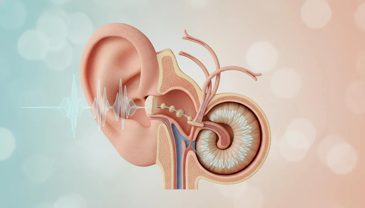
Every waking moment, your auditory system orchestrates one of the most mechanically elegant feats in human biology. Whether you are deciphering whispered speech across a crowded room, navigating traffic cues, or appreciating an orchestral symphony, your ears capture invisible atmospheric pressure waves and translate them into conscious neural reality in milliseconds. Yet, because hearing occurs effortlessly, few understand the delicate anatomical chain reactions required—or how vulnerable this pathway is to permanent damage.

Key Clinical Takeaways.
- Three-Part System: Auditory perception relies on the coordinated function of the outer ear (sound collection), middle ear (mechanical amplification), and inner ear (sensory transduction).
- Mechanical to Electrical: The cochlea contains about 15,000 delicate hair cells that convert hydraulic pressure waves into electrical signals via the vestibulocochlear nerve (Cranial Nerve VIII).
- Irreversible Loss: Human auditory hair cells cannot regenerate. Exposure to sounds exceeding 85 decibels (dBA) can cause permanent sensorineural hearing loss.
- Immediate Action for Sudden Changes: Unexplained sudden unilateral hearing loss is a medical emergency requiring rapid evaluation to prevent permanent deficit.
What Are the Anatomical Divisions of the Human Ear?
Clinically, the ear is divided into three distinct anatomical compartments, each serving a specialized functional phase of sound transmission: the outer ear, the middle ear, and the inner ear.
1. The Outer Ear: Acoustic Collection and Resonance.
The external auditory system functions as an acoustic antenna and funnel:
- The Auricle (Pinna): The cartilaginous framework visible externally. Its specialized convolutions (concha, helix, and antihelix) selectively filter and channel sound waves into the external auditory canal. It also provides spectral cues essential for vertical sound localization.
- External Auditory Canal: A 2.5 cm canal lined with epithelial tissue, fine hairs, and specialized ceruminous glands that produce cerumen (earwax). Cerumen maintains an acidic pH (about 6.0 to 6.5) providing natural antimicrobial defense and trapping foreign particulate matter. The canal also acts as an acoustic resonator, amplifying frequencies between 2,000 and 4,000 Hz—the critical range for human speech comprehension.
2. The Middle Ear: Impedance Matching and Mechanical Amplification.
The middle ear is an air-filled tympanic cavity housed within the temporal bone. Its primary task is overcoming "acoustic impedance mismatch"—the physical barrier between low-density air and high-density inner ear fluid.
- Tympanic Membrane (Eardrum): A taut, semi-transparent, three-layered membrane that separates the outer and middle ear. When airborne acoustic waves strike the membrane, it vibrates synchronously with their specific frequency and amplitude.
- The Auditory Ossicles: The body's three smallest bones form a connected lever system across the tympanic cavity:
- Malleus (Hammer): Attached directly to the inner surface of the tympanic membrane.
- Incus (Anvil): The intermediate bridge transmitting kinetic energy.
- Stapes (Stirrup): The smallest bone in the human body, whose footplate fits snugly into the oval window of the cochlea.
- Acoustic Amplification: Sound traveling straight from air into fluid would lose over 99% of its energy. The middle ear compensates using a mechanical lever advantage and an area ratio mechanism (the surface area of the eardrum is about 17 to 20 times larger than the stapes footplate), boosting acoustic pressure by roughly twenty-fold.
- Eustachian (Pharyngotympanic) Tube: Connects the middle ear to the nasopharynx. It periodically opens during swallowing and yawning to equalize middle ear pressure with atmospheric pressure, allowing optimal compliance of the tympanic membrane.
3. The Inner Ear: Fluid Dynamics and Sensory Transduction.
Deep within the petrous temporal bone lies the bony and membranous labyrinth, containing both vestibular (balance) organs and the primary organ of hearing:
- The Cochlea: A snail-shell-shaped organ spiraling roughly 2.5 to 2.75 turns, divided into three fluid-filled compartments: the scala vestibuli, scala media, and scala tympani.
- The Organ of Corti: The specialized sensory neuroepithelium resting on the basilar membrane. It houses rows of inner and outer hair cells capped by stereocilia.
- Tonotopic Organization: The basilar membrane is physically arranged like a piano keyboard. High-frequency sounds (up to 20,000 Hz) displace the narrow, stiff base near the oval window, while low-frequency sounds (down to 20 Hz) travel to the wide, flexible apex (helicotrema).
how it happens: How Mechanical Waves Become Neural Reality.
The transformation of sound into conscious thought follows a precise, sequential biophysical pathway:
- Acoustic Propagation: Pressure differentials in ambient air enter the external ear canal and set the tympanic membrane in motion.
- Mechanical Conduction: The malleus, incus, and stapes oscillate, transferring kinetic energy to the oval window.
- Hydraulic Displacement: In-and-out piston movements of the stapes footplate generate traveling waves through the perilymph and endolymph fluids within the cochlea.
- Stereocilia Shearing: Fluid motion deforms the basilar membrane, causing the stereocilia of hair cells to deflect against the overlying tectorial membrane.
- Electrochemical Depolarization: Mechanical deflection pulls microscopic tip links, opening mechanotransduction ion channels. Potassium (K^+) flows rapidly into hair cells from endolymph, depolarizing the cell membrane and opening voltage-gated calcium channels.
- Neurotransmitter Release: Synaptic vesicles release glutamate into the synaptic cleft, firing action potentials along the auditory branch of Cranial Nerve VIII (vestibulocochlear nerve).
- Central Auditory Processing: Signals ascend through the cochlear nucleus, superior olivary complex (enabling interaural time and intensity calculation for sound localization), inferior colliculus, and medial geniculate body of the thalamus, terminating in the primary auditory cortex (Brodmann areas 41 and 42) in the temporal lobe.
⚠️ Emergency Red Flags: When to Seek Immediate Medical Attention.
Certain auditory symptoms indicate serious conditions such as acute vascular compromise, temporal bone fracture, invasive infection, or stroke. Seek urgent assessment via 911 (US/Canada), 999 (UK), or your nearest emergency department (A&E / ER) if you experience:
- Sudden Sensorineural Hearing Loss (SSHL): Rapid hearing loss in one ear occurring within 72 hours, often noticed upon waking. This is an otologic emergency requiring early systemic corticosteroid therapy to preserve function.
- Neurological Deficits: Hearing impairment accompanied by facial weakness/droop, slurred speech, confusion, or limb numbness.
- Severe Vertigo and Incoordination: Inability to walk, intractable vomiting, or sudden visual disturbances alongside ear symptoms.
- Clear Fluid or Blood Otorrhea: Drainage of blood or clear cerebrospinal fluid (CSF) following head trauma.
- Periauricular Mastoiditis Signs: Erythema, warmth, swelling, or severe tenderness directly behind the ear accompanied by high fever.
Clinical Classification: Types of Hearing Loss.
Auditory impairment is classified according to the anatomical site of dysfunction:
| Condition Category. | Anatomical Site. / Common Etiologies. / Clinical Reversibility. |
|---|---|
| Conductive Hearing Loss. | Anatomical Site.: Outer or Middle Ear. Common Etiologies.: Cerumen impaction, otitis media with effusion, perforated tympanic membrane, otosclerosis. Clinical Reversibility.: often reversible via medical therapy or microsurgery. |
| Sensorineural Hearing Loss. | Anatomical Site.: Inner Ear (Cochlea) or Cranial Nerve VIII. Common Etiologies.: Presbycusis (age-related), chronic acoustic trauma, ototoxic drugs (aminoglycosides, cisplatin), viral labyrinthitis. Clinical Reversibility.: Typically permanent; managed with hearing amplification or cochlear implants. |
| Mixed Hearing Loss. | Anatomical Site.: Combined Conductive and Sensorineural. Common Etiologies.: Chronic suppurative otitis media with cochlear extension, head trauma, advanced otosclerosis. Clinical Reversibility.: Conductive component may resolve; sensorineural component is permanent. |
| Central Auditory Processing Disorder. | Anatomical Site.: Brainstem & Auditory Cortex. Common Etiologies.: Cerebrovascular events (stroke), traumatic brain injury, neurodegenerative disorders. Clinical Reversibility.: Variable; managed with auditory rehabilitation and specialized training. |
Protecting Your Auditory Health: Evidence-Based Do's & Don'ts.
Preventive otology focuses on protecting the delicate hair cells and maintaining the natural defense barriers of the ear canal.
| Clinical Recommendations (Do's). | Harmful Practices (Don'ts). |
|---|---|
| Practice the 60/60 Rule: Listen to personal audio at a maximum of 60% volume for no longer than 60 minutes at a time before resting your ears. | Never Insert Cotton Buds or Foreign Objects: Swabs, hairpins, and keys compress wax against the tympanic membrane and risk mechanical perforation. |
| Wear Hearing Protection: use custom earplugs or filtered ear defenders in noise environments exceeding 85 dBA (such as power tools, industrial work, and live music). | Avoid Unregulated "Ear Candling": This practice poses significant risks of burns, candle wax impaction, and tympanic membrane perforation without clinical efficacy. |
| Keep Ears Dry After Swimming: Gently tilt your head and towel-dry the outer ear to prevent maceration of the canal and otitis externa ("swimmer's ear"). | Do Not Ignore Early Tinnitus: Persistent ringing, buzzing, or hissing is an early warning sign of auditory distress that warrants audiological assessment. |
| Review medicines with Your Doctor: Inform your doctor of existing hearing issues before starting potentially ototoxic drugs (certain diuretics, antibiotics, or antineoplastics). | Avoid Delaying Evaluation of Sudden Loss: Waiting for spontaneous recovery in cases of sudden hearing loss can lead to permanent, irreversible sensory loss. |






No comments yet. Be the first to share your question or experience below.