

Consider the everyday precision required to fasten a small button, retrieve a coin from a smooth table, or type rapidly on a keyboard. Underpinning every delicate motion is your nail apparatus—an evolutionary marvel composed of specialized, compacted proteins designed to shield an exceptionally dense network of digital nerve endings. Far from being merely decorative keratin plates, human fingernails and toenails serve as mechanical countersinks that enhance fine tactile sensitivity while doubling as physiological barometers of systemic well-being.
Key Clinical Takeaways.
- Structural Composition: Nails consist of compacted, cross-linked, dead epithelial cells rich in hard alpha-keratin produced by the germinative nail matrix.
- Growth Velocity: Fingernails regenerate at an average physiological rate of 3.0 to 3.5 mm per month (requiring roughly 6 months for complete turnover), whereas toenails grow at about 1.0 to 1.6 mm per month (taking 12 to 18 months).
- Protective Barrier: The eponychium (cuticle) provides a hermetic antimicrobial seal protecting the germinal matrix; cutting or aggressively stripping it greatly increases the risk of acute and chronic paronychia.
- Clinical Window: Systemic conditions—including iron deficiency anemia, respiratory compromise, hepatic disease, and malignant melanoma—often manifest distinct diagnostic morphological signs across the nail unit.
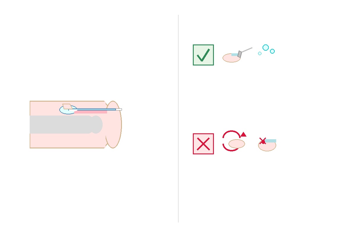
Anatomy of the Human Nail Apparatus: A Precision Biological Shield.
To clinical dermatologists, the human nail is known comprehensively as the onychostroma or nail unit. It comprises six mutually dependent anatomical structures, each executing a crucial biomechanical or protective role:
- The Germinal Matrix: Situated directly beneath the proximal nail fold, this specialized reproductive tissue generates the keratinocytes that form the hard nail plate. The proximal matrix forms the superficial two-thirds of the plate, while the distal matrix produces the inferior third. If this matrix suffers traumatic laceration, the nail plate can be permanently split or disfigured.
- The Lunula: The visible, pale, crescent-shaped arch at the proximal base of the nail. It represents the distal portion of the active germinal matrix. Its white appearance occurs because the underlying proliferative epithelial layer obscures underlying vascular capillaries.
- The Nail Plate: The rigid, translucent, keratinized shield that people casually identify as the "nail." It is composed of multiple compacted layers (dorsal, intermediate, and ventral) of dead, anucleated corneocytes bound by intercellular lipids.
- The Nail Bed (Sterile Matrix): The highly vascularized, sensory-nerve-rich connective tissue located directly beneath the adherent nail plate. It extends from the distal edge of the lunula to the hyponychium and anchors the advancing plate via longitudinal epidermal ridges.
- The Lateral and Proximal Nail Folds: The invaginations of vascularized digital skin that border and structurally stabilize the lateral and proximal borders of the nail plate.
- The Cuticle (Eponychium) and Hyponychium: Specialized cuticular seals. The proximal cuticle prevents water, pathogenic bacteria (such as Staphylococcus aureus), and dermatophytes from invading the proliferative matrix. The hyponychium seals the free distal edge underneath the fingertip, preventing onycholysis (separation of the nail plate from the bed).
how it happens and Growth: How Does Keratinization Work?
The progression from living cells to the durable, dead nail plate is governed by keratinization (specifically onychokeratinization). Stem cells within the germinal matrix continuously undergo active mitosis. As daughter cells synthesize high-sulfur fibrous keratin proteins, they are pushed upward and outward away from their basal nutrient and oxygen supplies.
During this migration, cellular organelles, nuclei, and cytoplasm condense and dissolve via programmed cellular condensation, leaving behind an exceptionally dense matrix of filamentous cross-linked proteins. Because these superficial cells are completely non-viable (dead), trimming the distal free edge causes zero physical pain. However, when trauma or invasive disease compromises the living tissue of the sterile nail bed or germinal matrix, intense sharp pain occurs due to the rich concentration of nociceptive nerve endings.
Clinical Signs: What Your Nails Reveal About Systemic Health.
Because the germinal matrix requires high metabolic turnover and sustained microvascular perfusion, systemic physiological disturbances often present visible abnormalities across the nail plate:
- Beau's Lines: Deep transverse horizontal depressions crossing the entire width of the nail plate. They arise from temporary arrest of cell division in the nail matrix during critical systemic insults, such as high-grade fevers, severe infections, myocardial infarction, or cytotoxic chemotherapy.
- Koilonychia ("Spoon Nails"): Concave fingernails where the lateral and distal margins flare upward, capable of holding a drop of water. This classic sign is most commonly correlated with chronic microcytic hypochromic iron deficiency anemia (often verified with serum ferritin < 30 ng/mL or < 30 mcg/L) and hemochromatosis.
- Digital Clubbing (Hippocratic Nails): An increase in the profile angle between the proximal nail fold and the nail plate (Lovibond's angle > 180°), accompanied by soft-tissue hypertrophy of the digital tuft. Clubbing often points to chronic hypoxemia, cardiopulmonary pathologies (such as bronchogenic carcinoma, chronic obstructive pulmonary disease, idiopathic pulmonary fibrosis, or infective endocarditis), and inflammatory bowel diseases.
- Terry's Nails: A clinical presentation where the proximal three-quarters of the nail appears pale, ground-glass white, with a narrow dark pink or brown transverse band at the distal tip. This sign reflects microvascular bed alterations secondary to end-stage hepatic cirrhosis, congestive heart failure, or severe malnutrition.
- Splinter Hemorrhages: Thin, linear, longitudinal red-to-brown marks under the nail plate. While minor physical trauma is the most frequent cause, multiple simultaneous splinter hemorrhages in non-traumatized digits raise immediate clinical suspicion for systemic lupus erythematosus, trichinellosis, or subacute bacterial endocarditis.
- Subungual Melanoma (Hutchinson's Sign): A persistent, widening longitudinal dark brown or black pigmented streak (longitudinal melanonychia) that spreads beyond the nail plate onto the adjacent proximal or lateral nail fold (Hutchinson's sign). This requires immediate histopathologic biopsy to rule out acral lentiginous melanoma.
🚨 Clinical Red Flags: When to Seek Urgent Medical Care.
Certain nail symptoms represent true dermatologic or vascular emergencies. Contact emergency medical services (call 911 in the United States/Canada or 999 in the United Kingdom) or proceed right away to the nearest Emergency Department (ER / A&E) if you notice:
- Peripheral Cyanosis: Rapid, acute blue or purplish discoloration of all nail beds combined with acute dyspnea, central chest pressure, altered mental status, or pulse oximetry falling below 90% (indicative of acute cardiopulmonary failure).
- Spreading Cellulitis or Ascending Lymphangitis: Severe, rapidly throbbing erythema, heat, and swelling surrounding a nail fold that streaks proximally up the finger, forearm, or wrist, accompanied by high fever (> 38.3°C / 101°F) or rigors.
- Deep Digital Pulp Abscess (Felon): Severe, tense, throbbing swelling of the palmar digital pulp space, threatening ischemic tissue necrosis of the phalanx.
Evidence-Based Nail Care: Clinical Best Practices.
Maintaining biological barrier integrity prevents invasive fungal infections (onychomycosis) and bacterial paronychia. Implement these validated dermatological standards:
| Clinical Best Practices (Do). | Harmful Practices (Avoid). |
|---|---|
| Trim Straight Across: Cut toenails cleanly straight across and gently smooth corner angles with an emery board to prevent jagged edges from embedding into the lateral folds (onychocryptosis or ingrown nail). | Do Not Round or Dig Down Corners: Tapering toenail margins into an oval shape drives the distal nail edge into the periungual tissue during ambulation, provoking painful ingrown nails. |
| Preserve the Cuticular Seal: Apply emollient ointments containing petroleum or ceramide compounds to maintain eponychial hydration and elasticity. | Do Not Excise or Forcefully Retract Cuticles: Mechanically clipping cuticles breaks the biological moisture seal, inviting acute paronychia from Pseudomonas or Candida albicans. |
| Practice Strict Digital Hygiene: Sanitize personal clippers with 70% isopropyl alcohol before and after each use; allow feet and shoes to dry thoroughly between wears. | Do Not Bite or Tear Hangnails: Onychophagia (chronic nail biting) and tearing hangnails strips protective skin layers, introducing oral flora into vascular spaces. |
doctor's Clinical Perspective.
"As clinicians, inspecting a patient's unpolished fingernails is often one of the first physical diagnostic steps we perform during a complete examination. The microvasculature beneath the nail plate provides real-time data on capillary refill, oxygen saturation, and nutritional adequacy. If a patient presents with an entirely detached nail from severe trauma, reassuring them is paramount: provided the germinal matrix remains viable and unscarred, a completely new, healthy fingernail will systematically replace itself over six to nine months. Treat your cuticles with care, maintain gentle hygiene, and bring any persistent, unexplained color or structural changes to your primary care doctor or board-certified dermatologist."
Scientific References & Authoritative Sources.
- National Health Service (NHS). Nail Problems and Fingernail Health: Overview and Red Flags. NHS England, 2024. Available at: nhs.uk/conditions/nail-problems.
- Fawcett RS, Linford S, Stulberg DL. Nail Abnormalities: Clues to Systemic Disease. American Family doctor (AAFP), 2004;69(6):1417-1424. Supported by clinical updates from Mayo Clinic Dermatology.
- Centers for Disease Control and Prevention (CDC). Nail Hygiene and Infection Prevention Standards. U.S. Department of Health and Human Services, 2023.
- National Institutes of Health (NIH) / National Library of Medicine. Anatomy and Physiology of the Nail Unit: Matrix, Bed, and Plate Dynamics. StatPearls Publishing, PubMed ID: NBK538289.


💬 Comments (0)
No comments yet. Be the first to share your thoughts here.
Add Your Comment