

That familiar, fiery burning sensation gnawing at your upper abdomen often gets brushed off as routine gastritis or simple indigestion after a heavy meal. Yet, when the discomfort persists, sharpens on an empty stomach, or wakes you in the dead of night, you might be dealing with something far more profound: a peptic ulcer eating away at your gastric lining. Much like a skin wound that struggles to heal under constant chemical exposure, a stomach ulcer represents an open sore on the delicate internal epithelium of your digestive tract. While modern medicine has rendered these lesions highly treatable, failing to recognize and manage them properly can lead to dangerous, life-threatening complications. Understanding the underlying how it happens, recognizing the cardinal warning signs, and pursuing evidence-based clinical interventions are vital steps toward protecting your digestive health.
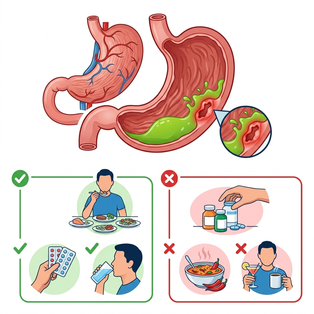
Navigating the complexities of digestive discomfort requires differentiating between transient irritation and structural pathology. In medical practice across leading global institutions like the NHS and Mayo Clinic, distinguishing between acute gastritis, gastroesophageal reflux disease (GERD), and true peptic ulcer disease (PUD) dictates the entire therapeutic strategy. This exhaustive clinical guide breaks down everything you need to know about stomach ulcers—from cellular mechanisms and diagnostic endoscopies to targeted medicine-based eradication therapies and long-term prevention protocols.
🔑 Key Clinical Takeaways.
- Primary Drivers: Over 80% of peptic ulcers are caused by chronic Helicobacter pylori bacterial infection or regular nonsteroidal anti-inflammatory drug (NSAID) use.
- Cardinal Symptom: A gnawing, burning epigastric pain located just below the sternum and above the umbilicus, often temporarily relieved or exacerbated by food intake.
- Diagnostic Gold Standard: Upper digestive endoscopy (EGD) allows direct visualization, tissue biopsy for H. pylori, and immediate therapeutic hemostasis if bleeding is present.
- Multimodal Treatment: Eradication requires combining proton pump inhibitors (PPIs) with targeted antibiotic regimens for bacterial infections or discontinuing ulcerogenic analgesics.
- Emergency Red Flags: Sudden, unbearable abdominal pain, black tarry stools (melena), or coffee-ground emesis demand immediate emergency medical intervention (ER / A&E / 911 / 999).
What is a Stomach Ulcer? (Overview).
How Do Stomach Ulcers Form?
To understand how a stomach ulcer develops, one must examine the hostile chemical environment inside the human stomach. The gastric mucosa secretes potent hydrochloric acid and digestive enzymes such as pepsin, which are essential for breaking down ingested food particles and neutralizing ingested pathogens. Because these substances are corrosive enough to dissolve organic tissue, the stomach relies on a sophisticated defense mechanism: a thick, continuous layer of bicarbonate-rich mucus that coats the epithelial lining, shielding it from autodigestion.
An ulcer forms when this protective mucosal barrier is compromised or when acid secretion overwhelms the tissue's defense capacity. Once the defensive mucous layer thins or breaks down, the highly acidic gastric juices make direct contact with the underlying epithelial cells. This caustic exposure creates an open sore, or erosion, that penetrates deeply through the mucosal muscularis layer. As gastric acid continuously irritates this raw tissue every time you eat or secrete digestive juices, the pain intensifies, and the body struggles to start natural wound repair without clinical intervention.
How Common Are Stomach Ulcers?
Peptic ulcer disease is a remarkably prevalent global health concern affecting millions of people annually. Its high frequency is directly linked to the widespread distribution of its two primary causative agents across modern populations:
- Unsupervised Analgesic Consumption: The widespread, frequent over-the-counter use of nonsteroidal anti-inflammatory drugs (NSAIDs) for chronic pain, headaches, and musculoskeletal disorders.
- Bacterial Colonization: High regional and global rates of chronic Helicobacter pylori bacterial infections residing undetected within the gastric mucosa.
Stomach Ulcers vs. Peptic Ulcers.
Patients often confuse terminology when discussing digestive sores. "Peptic ulcer disease" (PUD) serves as an overarching medical umbrella term describing any open sore formed within the mucosal lining of the stomach (gastric ulcer) or the upper section of the small intestine known as the duodenum (duodenal ulcer). so, every stomach ulcer is classified as a peptic ulcer. This duodenal ulcers occur just past the pyloric sphincter. Although their exact anatomical locations differ slightly, both conditions share remarkably similar underlying body mechanisms, diagnostic pathways, and therapeutic regimens.
Symptoms and Causes.
What Are the Symptoms of a Stomach Ulcer?
- Cardinal Symptom: Epigastric Burning and Pain: The hallmark clinical presentation of a gastric ulcer is a distinct burning sensation or gnawing pain localized in the upper central abdomen—specifically beneath the xiphoid process and above the umbilicus, often shifting slightly to the left. Patients often describe this sensation as feeling like acid is burning the tissue or an internal gnawing ache. This pain stems directly from gastric acid and digestive enzymes irritating the exposed nerve endings within the open mucosal crater. The discomfort often peaks when the stomach is empty—between meals or during the middle of the night—when acid secretion continues without buffering food particles. While duodenal ulcer pain is classically relieved by eating, gastric ulcer pain may sometimes worsen shortly after food ingestion as gastric acid production surges.
- Dyspepsia and Postprandial Fullness: people with active stomach ulcers routinely experience chronic dyspepsia. This encompasses persistent upper abdominal discomfort accompanied by early satiety (feeling uncomfortably full shortly after starting a meal) or prolonged postprandial fullness where food seems to linger sluggishly in the upper GI tract.
-
Other Common Associated signs: Depending on secondary inflammation and person physiological variations, patients may report:
- Gastroesophageal reflux (acid reflux) and retrosternal heartburn .
- Persistent nausea and occasional episodes of non-bloody vomiting .
- Abdominal bloating, frequent eructation (belching), and painful gas accumulation .
-
Silent Ulcers and Complication Warnings: A significant clinical challenge is the existence of "silent ulcers," wherein patients harbor active, bleeding, or deep mucosal lesions without having typical pain or dyspepsia. In these cases, the condition remains entirely asymptomatic until a catastrophic medical event—such as acute digestive hemorrhage or visceral perforation—occurs. Seek urgent medical evaluation right away if you experience any of the following red-flag symptoms:
- Notable cutaneous pallor or pale nail beds .
- Unexplained dizziness, profound physical weakness, or presyncopal lightheadedness, which often indicate acute or chronic anemia .
- Dark, tarry, foul-smelling stools (melena), signaling upper digestive bleeding where blood has been digested along the intestinal tract .
- Vomiting material that resembles dark coffee grounds, indicating oxidized blood mixed with gastric contents .
- Sudden, severe, unrelenting abdominal pain that rapidly intensifies, suggesting acute visceral perforation .
How Serious Are Complications?
Unchecked or inadequately treated peptic ulcers can precipitate severe, life-threatening medical emergencies:
- Bleeding Ulcers: Chronic oozing or sudden erosion into a major submucosal blood vessel can cause continuous or massive hemorrhage. Slow, chronic blood loss leads inexorably to iron-deficiency anemia, causing fatigue and shortness of breath. Acute massive hemorrhage induces hypovolemic shock, presenting a critical medical emergency requiring immediate resuscitation.
- Perforated Ulcers: When an ulcer completely erodes through the full thickness of the gastric or duodenal wall, gastric contents, acid, and bacteria spill freely into the sterile peritoneal cavity. This precipitates chemical and bacterial peritonitis—a severe, agonizing abdominal infection that can rapidly transition into systemic sepsis and multi-organ failure.
What Are the Main Causes of Stomach Ulcers?
Global clinical research confirms that the overwhelming majority of stomach ulcers stem from two primary etiologies:
- Helicobacter pylori Bacterial Infection: H. pylori is a spiral-shaped, microaerophilic bacterium capable of surviving the highly acidic human stomach by producing the enzyme urease, which neutralizes local acid. Roughly half of the global population harbors this bacterium, often acquired during childhood. While many infected people remain asymptomatic, certain strains trigger persistent chronic inflammation (chronic active gastritis) by degrading the protective mucous layer and provoking localized immune responses. Over years, this chronic inflammatory cascade damages the epithelial barrier, culminating in gastric or duodenal ulceration.
- Nonsteroidal Anti-Inflammatory Drug (NSAID) Usage: Regular, long-term, or high-dose consumption of nonsteroidal anti-inflammatory drugs represents the second major catalyst for peptic ulcer disease. Commonly utilized analgesics such as ibuprofen, naproxen, diclofenac, and prescription aspirin (notably distinct from paracetamol/acetaminophen, which lacks systemic anti-inflammatory properties) directly irritate the gastric mucosa. also, NSAIDs systemically inhibit cyclooxygenase-1 (COX-1) enzymes, blocking the synthesis of cytoprotective prostaglandins. These vital prostaglandins normally stimulate bicarbonate and mucus secretion while maintaining mucosal blood flow. Suppressing them leaves the gastric lining defenseless against acid erosion.
Other Rarer Causes.
Beyond H. pylori and NSAIDs, select uncommon medical conditions can trigger ulcer formation:
- Rare Pathogenic Infections: Exceptional bacterial, viral, or fungal infections impacting the upper digestive tract.
- Zollinger-Ellison Syndrome: A rare neuroendocrine tumor syndrome characterized by gastrin-secreting tumors (gastrinomas) located in the pancreas or duodenum. Excessive circulating gastrin drives relentless, hyper-secretory gastric acid production, overwhelming mucosal defenses and causing refractory, multiple peptic ulcers.
- Severe Physiological Stress ("Stress Ulcers"): Critical illness, extensive thermal burns (Curling's ulcers), severe intracranial trauma (Cushing's ulcers), or major emergency surgeries induce profound physiological stress. The resulting visceral hypoperfusion and neuroendocrine shifts compromise mucosal integrity, predisposing vulnerable hospitalized patients to acute stress-related mucosal damage. Note that this physiological phenomenon is entirely distinct from everyday emotional or psychological stress.
Does Lifestyle Play a Role?
Popular myth often attributes stomach ulcers directly to eating spicy foods or having high psychological stress. Clinically speaking, routine daily lifestyle factors (such as dietary spices or everyday anxiety) do not directly start stomach ulcers. However, if an active ulcer is already present, these factors can greatly worsen clinical symptoms by stimulating gastric acid secretion and further irritating the exposed mucosal lesion:
- Cigarette smoking (which impairs mucosal healing and blood flow) .
- Excessive alcohol consumption .
- Spicy, capsaicin-rich foods .
- Highly acidic foods (e.g., citrus fruits, tomatoes) .
- Caffeine-heavy beverages such as coffee and strong black tea .
Diagnosis and Tests.
How is a Stomach Ulcer Diagnosed?
When presenting with persistent upper abdominal discomfort, a doctor begins with a thorough medical history review. This evaluation focuses on symptom chronology, the frequency of over-the-counter analgesic consumption, and any prior history of digestive illness or H. pylori exposure. If clinical suspicion of peptic ulcer disease is high, objective diagnostic testing is required to visualize the lesion and confirm its underlying etiology.
What Kinds of Tests Are Performed?
Clinicians generally seek answers to two pivotal questions during the diagnostic workup:
- Does the patient harbor an active H. pylori bacterial infection?
- Is there a structural mucosal ulcer present, and what are its precise morphological characteristics?
Both objectives are often achieved through a single clear investigation: upper digestive endoscopy.
- Upper digestive Endoscopy (EGD): Considered the clinical gold standard for evaluating upper digestive pathology, an esophagogastroduodenoscopy involves passing a flexible, illuminated fiber-optic scope (endoscope) through the oral cavity, down the esophagus, and into the stomach and proximal duodenum under light procedural sedation. This allows the gastroenterologist to directly visualize the gastric mucosa in real-time on high-definition monitors, accurately identifying mucosal breaks, active bleeding sites, or structural irregularities. During the procedure, specialized miniature instruments can be passed through the working channel to harvest tiny mucosal tissue samples (biopsies) for rapid urease testing or histological examination confirming H. pylori. also, if an ulcer is actively bleeding, therapeutic interventions—such as electrocautery, thermal coagulation, or hemostatic clips—can be applied right away during the same session to arrest hemorrhage.
-
Alternative Diagnostic Investigations: While EGD remains superior, alternative or adjunctive evaluations include:
- Barium Swallow (Upper GI Series): The patient swallows a radiopaque barium suspension while serial fluoroscopic X-rays are captured. Barium coats the mucosal contour, outlining craters and filling ulcerative niches. Although non-invasive, this technique possesses lower sensitivity than endoscopy and cannot detect superficial erosions or obtain tissue biopsies.
-
H. pylori Screening Modalities:
- Urea Breath Test: The patient ingests a labeled carbon isotope solution; if active H. pylori urease is present, it metabolizes the compound, releasing labeled carbon dioxide detected in exhaled breath samples.
- Serological Blood Tests: Detect circulating anti-H. pylori antibodies. However, because antibodies persist long after successful bacterial eradication, this test cannot distinguish between current active infections and past resolved exposures.
- Stool Antigen Test: Analyzes a stool sample for specific bacterial proteins, confirming active digestive colonization.
Conditions That Can Mimic a Stomach Ulcer.
If diagnostic endoscopies or imaging studies rule out a peptic ulcer, persistent symptoms may stem from alternative digestive disorders:
- Gastroesophageal Reflux Disease (GERD): Chronic retrograde flow of acidic gastric contents into the esophagus, producing retrosternal heartburn and acid regurgitation.
- Functional Dyspepsia: A common non-ulcer dyspepsia characterized by chronic, bothersome upper abdominal fullness, early satiety, or epigastric pain without any clear structural lesion or mucosal break on endoscopy.
- Gastric Malignancy (Stomach Cancer): Although statistically less common, gastric carcinoma can present with ulcerated lesions mimicking benign peptic ulcers (e.g., persistent pain, early satiety, unexplained weight loss). This underscores why endoscopic biopsy is mandatory during evaluation.
| Condition. | Primary Mechanism. | Classic Symptom Pattern. | Preferred Diagnostic Test. |
|---|---|---|---|
| Peptic / Stomach Ulcer. | Mucosal barrier breakdown from H. pylori or NSAIDs. | Gnawing, burning epigastric pain, often worse when fasting. | Upper Endoscopy (EGD) with biopsy. |
| Gastroesophageal Reflux (GERD). | Lower esophageal sphincter dysfunction. | Retrosternal heartburn, acid regurgitation, sour taste. | Clinical evaluation / 24-hr pH monitoring. |
| Functional Dyspepsia. | Visceral hypersensitivity and motility dysfunction. | Postprandial fullness, early satiety, upper discomfort. | EGD (to exclude organic disease). |
Management and Treatment.
How to Heal a Stomach Ulcer Promptly.
Accelerating the healing of a gastric ulcer requires systematically dismantling the underlying pathogenic driver. For instance, if diagnostic findings implicate NSAID use, right away discontinuing the offending analgesic often allows the mucosal tissue to heal naturally, even without aggressive pharmacotherapy. Conversely, if H. pylori colonization is confirmed, successfully eradicating the bacteria with targeted multi-antibiotic regimens is non-negotiable. doctors supplement these root-cause interventions with antisecretory medicines that suppress gastric acid output, providing a protective, low-acid microenvironment where epithelial regeneration can proceed rapidly.
What Treatments Are Available?
Modern therapeutic protocols for peptic ulcer disease rely on combination pharmacotherapy designed to suppress acid production, fortify mucosal defenses, and eradicate bacterial pathogens.
-
medicine-based Interventions:
- Antibiotic Regimens: When H. pylori is detected, standard eradication therapy employs a multi-drug protocol combining a proton pump inhibitor with two or three specific antibiotics (commonly including clarithromycin, amoxicillin, metronidazole, or tetracycline) given concurrently for 7 to 14 days. Patients must complete the entire prescribed course without interruption to prevent treatment failure and antibiotic resistance.
- Cytoprotective Agents: medicines that form a protective physical coating over the ulcer base or stimulate endogenous prostaglandin synthesis to bolster mucosal defense. Common examples include sucralfate, misoprostol, and bismuth subsalicylate. These are especially valuable in managing and preventing NSAID-induced mucosal injury.
- H2 Receptor Blockers (H2 Blockers): Competitive antagonists of histamine at the parietal cell H2 receptors, reducing basal and stimulated gastric acid secretion. Examples include famotidine, cimetidine, and nizatidine.
- Proton Pump Inhibitors (PPIs): Potent suppressors of gastric acid production that irreversibly bind to the hydrogen-potassium ATPase pump (the proton pump) on gastric parietal cells. PPIs represent the cornerstone of modern ulcer therapy, rapidly relieving symptoms and fostering high ulcer healing rates. Commonly prescribed agents include omeprazole, esomeprazole, lansoprazole, pantoprazole, and rabeprazole.
-
Endoscopic and Surgical Management:
- Endoscopic Hemostasis: For actively bleeding ulcers discovered during EGD, gastroenterologists use endoscopic thermal coagulation, hemoclips, or sclerotherapy injections to secure the bleeding vessel and achieve hemostasis.
- Emergency Surgery for Perforation: Visceral perforation requires emergency exploratory laparoscopy or laparotomy to close the perforated defect and thoroughly lavage the peritoneal cavity.
- Surgical Interventions for Refractory Disease: Though exceptionally rare in the era of modern pharmacotherapy, chronic refractory ulcers or severe scarring causing gastric outlet obstruction may require surgical procedures such as pyloroplasty (widening the pyloric channel) or selective vagotomy (cutting branches of the vagus nerve to permanently reduce acid output).
Prevention.
How to Prevent or Prevent Recurrence of Stomach Ulcers.
Preventing initial ulcer development or stopping recurrent episodes involves proactive lifestyle and clinical risk management:
- Eradicating H. pylori: Screening and treating asymptomatic or symptomatic H. pylori colonization eliminates chronic mucosal inflammation before ulcer craters can form.
- Judicial NSAID Administration: Patients requiring chronic anti-inflammatory therapy for conditions like osteoarthritis should collaborate with their doctors to use the lowest effective analgesic dose for the shortest possible duration. When long-term NSAID use is unavoidable, co-prescribing a prophylactic proton pump inhibitor or switching to a COX-2 selective inhibitor can greatly reduce digestive toxicity.
- Modulating Lifestyle Risk Factors: Eliminating tobacco smoking and moderating alcohol consumption removes potent mucosal irritants that impair epithelial repair mechanisms.
Outlook / Prognosis.
How Long Does It Take for a Stomach Ulcer to Heal?
When patients strictly adhere to prescribed antisecretory pharmacotherapy, complete H. pylori eradication courses, and discontinue ulcerogenic medicines (such as NSAIDs), uncomplicated gastric ulcers typically heal completely within several weeks (generally 4 to 8 weeks). Clinicians often schedule follow-up evaluations or repeat endoscopies to confirm total mucosal healing and ensure bacterial eradication.
Chronic Conditions.
While most people require only short-term treatment courses, patients harboring rare chronic hypersecretory syndromes like Zollinger-Ellison syndrome require long-term, ongoing antisecretory management to maintain remission and prevent recurrent ulceration.
Living With.
The Importance of Seeking Professional Medical Advice.
If you suspect you are developing a stomach ulcer, seeking professional medical evaluation is essential. Over-the-counter antacids or bismuth subsalicylate preparations can neutralize acid and offer comforting temporary relief, yet they do nothing to heal the underlying mucosal defect. True healing requires identifying and treating the root biological cause—whether that entails eradicating H. pylori or adjusting analgesic regimens.
Risks of Leaving Ulcers Untreated.
Even in the absence of excruciating pain, leaving a peptic ulcer untreated exposes the patient to severe cumulative risks, including silent digestive bleeding, chronic iron-deficiency anemia, visceral perforation, peritonitis, and an elevated long-term epidemiological risk of gastric malignancy associated with chronic untreated H. pylori infection.
When to Seek Emergency Medical Care.
Proceed right away to the nearest Emergency Department (ED / A&E / ETU) or call emergency services (911 in the US/Canada, 999 in the UK) if you experience any of the following emergency indicators:
- Sudden, excruciating, unrelenting abdominal pain that spreads rapidly across the abdomen .
- Passing dark, tarry stools or vomiting bright red blood or dark coffee-ground emesis .
- Profound systemic symptoms of acute blood loss, including severe dizziness, fainting, clammy skin, and profound weakness .
🩺 A Clinical Note from Nirogi Lanka.
Chronic upper abdominal pain should never be casually dismissed as routine gastritis without proper diagnostic verification. Modern gastroenterology offers precise, highly effective tools—ranging from non-invasive breath tests to advanced high-definition endoscopies—that can identify the exact root cause of your discomfort. By partnering with your doctor to address bacterial infections or modify analgesic habits, you can achieve permanent relief, restore mucosal integrity, and protect your long-term digestive health.


💬 Comments (0)
No comments yet. Be the first to share your thoughts here.
Add Your Comment