

When your doctor tells you to have a breast ultrasound , you may be a little scared and curious. 'What is it?', 'Will it hurt?', 'Why did you ask me to do this?' You probably have a lot of questions in your mind, right? Don't be afraid of anything. This is actually a much simpler and less painful test than you think. Today, we will talk about this Breast Ultrasound in a very simple and friendly way.
What is a breast ultrasound?
Simply put, it's a painless test, similar to a prenatal ultrasound scan, that uses high-frequency sound waves to create an image or video of the tissue inside your breast on a computer screen.
Just like when we walk up a hill and talk, we hear an echo when we hit it, the sound waves emitted by this scanning device bounce off the various tissues inside the breast and come back. The echoes that come back are analyzed by a computer and converted into a clear image. This is a very safe test because it does not emit any radiation into the body like X-rays.
Why are we being asked to do this scan?
There are several reasons why a doctor might ask you to have this test. It's not always something to be afraid of. Sometimes it's done as a further confirmation. Let's look at the main reasons.
| Reason for referral for testing | A simple explanation |
|---|---|
| If the breast tissue density is high | Some women have naturally dense breast tissue. In such cases, it can be difficult to get a clear image from a mammogram. Ultrasound can clearly see through this dense tissue. |
| If you are pregnant | Since unnecessary radiation exposure is not advisable during pregnancy, a safer ultrasound scan is recommended instead of a mammogram. |
| If you are under 25 years old | Young women typically have denser breast tissue, so an ultrasound is often the first test of choice when they have a breast problem. |
| If there is a suspicious area on a mammogram | If a mammogram identifies a suspicious area, it is examined further, which means using an ultrasound, as if zooming in. |
| Check to see if a lump is a cyst or solid. | This scan can accurately distinguish and identify whether a lump in the breast is a harmless fluid-filled bubble (cyst), or a solid mass made of tissue (solid). This is very important. |
| As an aid to a biopsy | If you need to take a piece of tissue (biopsy) from a breast lump, ultrasound is used to guide the needle to the exact location of the lump, like a GPS. |
How should I prepare before the scan?
This doesn't require much preparation. It's very simple.
- Your doctor will explain the test to you. Ask any questions you may have at that time.
- You can eat and drink normally before the scan . There is no need to fast.
- The most important thing is that on the morning of the test, do not apply any lotion, powder, cream, deodorant, or perfume to your breasts and armpits, as some of these substances can affect the clarity of the scan image.
- Wear clothes that are easy to remove. Usually, you will have to expose your upper body from the waist down. Don't worry about that, the hospital will give you a special gown to wear during that time.
How is the scan done?
If you know what will happen step by step when you go into the scan room, your fear will be reduced even more.
1. First, you will be asked to lie on your back on an examination table. You may also be asked to place your hands above your head.
2. Then, the technician will apply a small amount of water-soluble gel to the area of your breast that will be scanned. This will feel a little cold, but it won't hurt your skin. If it gets on your clothes, you can wash them without leaving any stains.
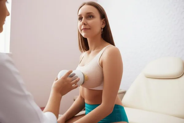
3. Next, a device (probe) that looks like a small microphone or a small bridge is slowly moved back and forth over the gel- coated skin. This is not painful. It may feel like a slight pinch.
4. Sometimes you will be asked to hold your breath for a while to get a clear image.
5. The entire test only takes between 10 and 20 minutes .
6. When the test is finished, the applied gel will be wiped off with a tissue. You can then get dressed and continue your normal activities without any problems.
How can I find out about my report (Results)?
A radiologist (radiologist) examines the images from the scan and prepares a report. He or she will forward the report to the doctor who is treating you.
Your doctor will review the report and explain it to you in a simple and clear manner. So you should definitely see your doctor to discuss the results.
You may need further tests. This may be due to:
- If the doctor thinks that a problematic area needs to be examined more closely or through another test,
- Observe if any changes occur over time.
- If you are undergoing any treatment, check to see if it is effective.
Take-Home Message
- Breast Ultrasound is a painless, very safe scan that uses sound waves.
- It is commonly used to look for a lump in the breast, further examine a suspicious area on a mammogram, and guide a biopsy.
- Do not apply any lotion, cream, powder, or deodorant to the breast area on the day of the test.
- Your doctor will explain the results of the scan to you. Don't be afraid to ask him any questions you have.




💬 Comments (0)
No comments yet. Be the first to share your thoughts here.
Add Your Comment